. 2013;8(1):e54747.
doi: 10.1371/journal.pone.0054747. Epub 2013 Jan 24.

Spindle shaped human mesenchymal stem/stromal cells from amniotic fluid promote neovascularization

Affiliations

Spindle shaped human mesenchymal stem/stromal cells from amniotic fluid promote neovascularization

Maria G Roubelakis et al. PLoS One. 2013.

Abstract

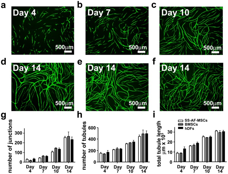
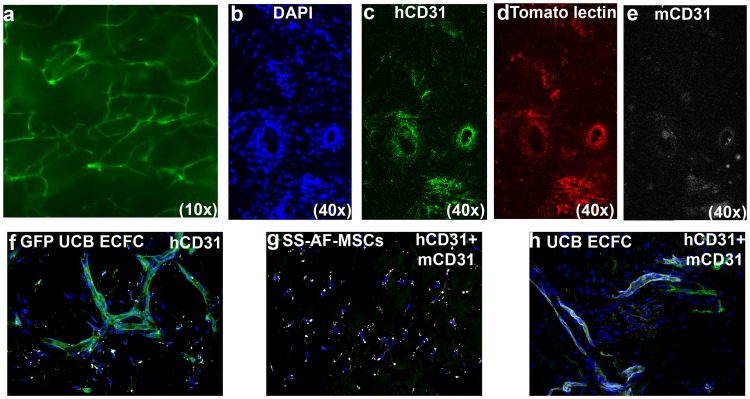
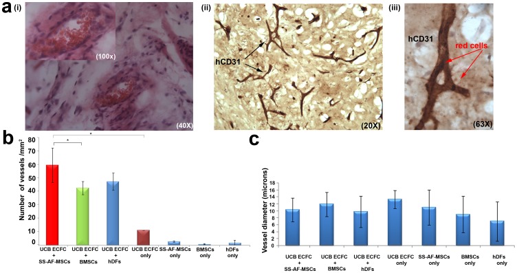
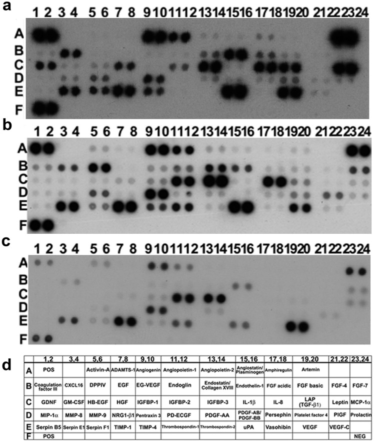
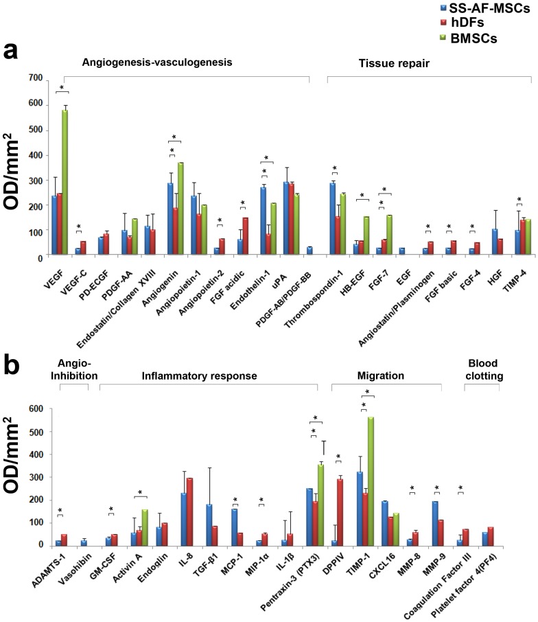
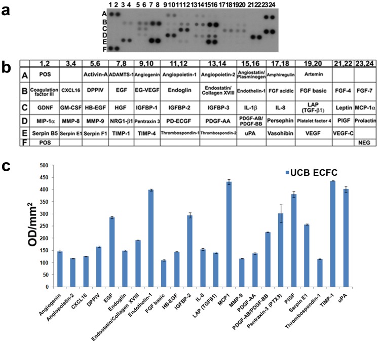
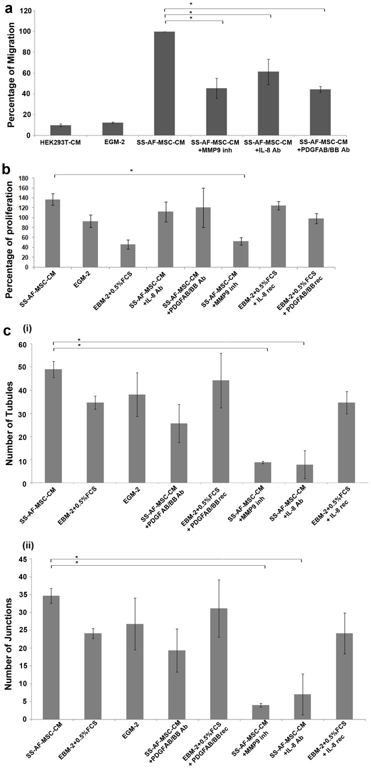
Human amniotic fluid obtained at amniocentesis, when cultured, generates at least two morphologically distinct mesenchymal stem/stromal cell (MSC) subsets. Of these, the spindle shaped amniotic fluid MSCs (SS-AF-MSCs) contain multipotent cells with enhanced adipogenic, osteogenic and chondrogenic capacity. Here, we demonstrate, for the first time, the capacity of these SS-AF-MSCs to support neovascularization by umbilical cord blood (UCB) endothelial colony forming cell (ECFC) derived cells in both in vitro and in vivo models. Interestingly, although the kinetics of vascular tubule formation in vitro were similar when the supporting SS-AF-MSCs were compared with the best vasculogenic supportive batches of bone marrow MSCs (BMSCs) or human dermal fibroblasts (hDFs), SS-AF-MSCs supported vascular tubule formation in vivo more effectively than BMSCs. In NOD/SCID mice, the human vessels inosculated with murine vessels demonstrating their functionality. Proteome profiler array analyses revealed both common and distinct secretion profiles of angiogenic factors by the SS-AF-MSCs as opposed to the hDFs and BMSCs. Thus, SS-AF-MSCs, which are considered to be less mature developmentally than adult BMSCs, and intermediate between adult and embryonic stem cells in their potentiality, have the additional and very interesting potential of supporting increased neovascularisation, further enhancing their promise as vehicles for tissue repair and regeneration.


Conflict of interest statement

Competing Interests: The authors have declared that no competing interests exist.

Figures

Figure 1
Figure 1. SS-AF-MSCs support neovascularisation in vitro – comparing the dynamics of vessel formation in the presence of SS-AF-MSCs, hDFs or BMSCs.
(a-f) Representative fields of vascular tubules after (a) 4, (b) 7, (c) 10, and (d to f) 14 days of co-culture of eGFP UCB ECFC derived cells with (a to d) SS-AF-MSCs, and pre-selected optimized batches of (e) BMSCs or (f) hDFs respectively. Scale bar = 500 µm. (g-i) Quantification of vascular tubule phenotypes at days 4, 7, 10 and 14, showing no significant difference in the number of junctions, tubules and total tubule length during the 14 days of co-culture between the 3 stromal cell types (p>0.05 Student’s t test). Values are means±S.D. for three independent experiments.
Figure 2
Figure 2. Immunofluorescence vessel imaging.
(a) Representative photomicrographs of the matrigel implants containing eGFP-UCB ECFC derived cells and SS-AF-MSCs. (b-e) Representative photomicrographs of matrigel implant sections containing non eGFP tagged UCB ECFC derived cells and SS-AF-MSCs after staining with (b) DAPI (blue), (c) hCD31 (green), (d) following tomato lectin perfusion (red), and (e) mCD31 (white). (f) Co-localization of eGFP (green) with hCD31 (white) staining in matrigel implant sections containing eGFP-UCB ECFC derived cells and SS-AF-MSCs. (g) Representative photomicrographs of matrigel implant sections containing SS-AF-MSCs only stained for hCD31 (green) and mCD31 (white) antigens, but where hCD31 was not detected. (h) Representative photomicrographs of matrigel implant sections containing UCB ECFC derived cells only, stained for hCD31 (green) and mCD31 (white) antigens.
Figure 3
Figure 3. Quantitating vessel formation in in vivo studies.
(a) Histological evaluation of vessels containing SS-AF-MSCs and UCB ECFC derived cells, harvested 14 days post-implantation and stained (i) with hematoxylin/eosin and for (ii-iii) human CD31 antigen (brown stain). High-power view of a vessel containing red blood cells (arrowed) from (ii) is shown in (iii). (b) Microvessel density in matrigel implants containing combined SS-AF-MSCs, BMSCs or hDFs with UCB ECFC derived cells, SS-AF-MSCs only, BMSCs only, hDFs only or UCB ECFC derived cells only. Vessel number (vessels/mm2) was estimated using Image J 1.38× software. Statistical analysis was performed using Student’s t test. (c) Vessel diameter estimation in matrigel implants containing SS-AF-MSCs, BMSCs or hDFs and UCB ECFC derived cells, SS-AF-MSCs only, or UCB ECFC derived cells only using Image J 1.38× software, (*p<0.05 Student’s t test). A minimum of 15 fields of view (40x) were analyzed from each photograph. Error bars indicate S.D. of the mean for 10 photographs from each group.
Figure 4
Figure 4. Analysis of angiogenic factors secreted by SS-AF-MSCs, BM-MSCs and hDFs in vitro using proteome arrays.
(a-c) Representative proteome profiler arrays for (a) SS-AF-MSCs, (b) BMSCs and (c) hDFs respectively; (d) corresponding names of each molecule within the array summarized in tabular form.
Figure 5
Figure 5. Secreted angiogenic factors in SS-AF-MSC-, BMSCs and hDF- conditioned media (CM) using proteome arrays.
(a, b) Relative expression levels of angiogenic factors secreted from SS-AF-MSC-, BMSC- and hDF-CM according to their function. Values are normalized to positive controls. Values are means ± S.D. for three independent experiments, (*p<0.05 Student’s t test).
Figure 6
Figure 6. Analysis of angiogenic factors secreted by UCB ECFC derived cells in vitro.
(a) Representative proteome profiler array for UCB ECFC derived cell-CM; (b) corresponding names of each molecule within the array summarized in tabular form; (c) Relative expression levels of angiogenic factors in UCB ECFC derived cell-CM. Values are normalized to positive controls. Values are means ± S.D. for three independent experiments, (*p<0.05 Student’s t test).
Figure 7
Figure 7. Role of IL-8, PDGF-AB/BB and MMP9 molecules from conditioned media in migration, proliferation and ability of tubule formation of UCB ECFC derived cells.
(a) Histograms showing the migration of UCB ECFC derived cells towards SS-AF-MSC-conditioned medium (CM), EGM-2 medium, control medium (EBM-2, 0.5% (v/v) FCS), SS-AF-MSC-CM+IL8 neutralizing Ab, SS-AF-MSC-CM +PDGF-AB/BB neutralizing Ab or SS-AF-MSC-CM+MMP9 inhibitor (inh). Values are means ± S.D. for three independent experiments (*p<0.05 Student’s t test). (b) Examination of the proliferation rate in vitro of UCB ECFC derived cells under the same conditions. Control medium with recombinant (rec) IL-8 or PDGF-AB/BB was also included. Values are means ± S.D. for three independent experiments, (*p<0.05 Student’s t test). (c) In vitro angiogenesis matrigel assay for UCB ECFC derived cells under the respective conditions for estimation of the number of (i) tubules and (ii) junctions formed. Error bars indicate S.D. of the mean for 10 (5x) photographs from each group (*p<0.05 Student’s t test).

References

    1. Friedenstein AJ, Chailakhyan RK, Latsinik NV, Panasyuk AF, Keiliss-Borok IV (1974) Stromal cells responsible for transferring the microenvironment of the hemopoietic tissues. Cloning in vitro and retransplantation in vivo. Transplantation 17: 331–340. - PubMed
    1. Caplan AI (1991) Mesenchymal stem cells. J Orthop Res 9: 641–650. - PubMed
    1. Pittenger MF, Mackay AM, Beck SC, Jaiswal RK, Douglas R, et al. (1999) Multilineage potential of adult human mesenchymal stem cells. Science 284: 143–147. - PubMed
    1. Dexter TM, Allen TD, Lajtha LG (1977) Conditions controlling the proliferation of haemopoietic stem cells in vitro. J Cell Physiol 91: 335–344. - PubMed
    1. English K, French A, Wood KJ (2011) Mesenchymal stromal cells: facilitators of successful transplantation? Cell Stem Cell 7: 431–442. - PubMed

Publication types

Substances