Review
. 2013 Feb;32(1):4-18.
doi: 10.3109/08830185.2012.749400.

Host defense at the ocular surface

Affiliations
Review

Host defense at the ocular surface

Eric Pearlman et al. Int Rev Immunol. 2013 Feb.

Abstract

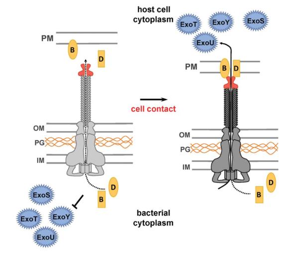
Microbial infections of the cornea frequently cause painful, blinding and debilitating disease that is often difficult to treat and may require corneal transplantation. In addition, sterile corneal infiltrates that are associated with contact lens wear cause pain, visual impairment and photophobia. In this article, we review the role of Toll-Like Receptors (TLR) in bacterial keratitis and sterile corneal infiltrates, and describe the role of MD-2 regulation in LPS responsiveness by corneal epithelial cells. We conclude that both live bacteria and bacterial products activate Toll-Like Receptors in the cornea, which leads to chemokine production and neutrophil recruitment to the corneal stroma. While neutrophils are essential for bacterial killing, they also cause tissue damage that results in loss of corneal clarity. These disparate outcomes, therefore, represent a spectrum of disease severity based on this pathway, and further indicate that targeting the TLR pathway is a feasible approach to treating inflammation caused by live bacteria and microbial products. Further, as the P. aeruginosa type III secretion system (T3SS) also plays a critical role in disease pathogenesis by inducing neutrophil apoptosis and facilitating bacterial growth in the cornea, T3SS exotoxins are additional targets for therapy for P. aeruginosa keratitis.


Figures

FIGURE 1
FIGURE 1. Resident myeloid cells in the cornea
A,B: CX3CR1GFP+ cells in a normal mouse cornea (A, original magnification is x20), and the distribution of epithelial and stromal dendritic cells (DC) and stromal macrophages (B). C: Mouse corneal whole mounts from wild type (panel 1, 4), CD11c eYFP (panel 3) or CX3CR1GFP+ mice were incubated with antibodies to MHC class II or CD11b, counterstained with DAPI, and visualized by confocal microscopy. Images of each cell population illustrated in Figure 1B is shown in panels 1–4, including in vivo MHC II membrane nanotubes (panel 4), which were present in the cornea 24h after topical exposure to+LPS. Original magnification is x400, and similar images and detailed descriptions are in references [35, 39, 40].
FIGURE 2
FIGURE 2. Predicted sequence of events in P. aeruginosa keratitis
Following corneal abrasion and infection, TLR4/MD-2 and TLR5 are activated on resident macrophages leading to production of CXC chemokines and pro-inflammatory cytokines by resident macrophages () including IL-1α and IL-1β. These cytokines mediate neutrophil (PMN) recruitment from peripheral capillaries in the cornea, which secrete reactive oxygen and nitrogen species (ROS, RNS) and matrix metalloproteinases (MMP) that disrupt the normal architecture of the corneal stroma and results in loss of corneal clarity. IL-1α and IL-1β have autocrine and paracrine effects on cells expressing IL-1R1, including corneal epithelial cells (HCEC), resulting in production of chemokines and antimicrobial peptides (AMP), which exacerbate the inflammatory response. The diagram is based primarily from data in reference [40].
FIGURE 3
FIGURE 3. LPS responsiveness in human corneal epithelial (HCE) cells
Following infection with P. aeruginosa, natural killer (NK) cells are recruited to the corneal stroma, and produce IFN-γ. IFN-γ receptor activation on corneal epithelial cells (epi) leads through p-STAT1 to MD-2 transcription and translocation to the cell surface with constitutively expressed TLR4. Corneal epithelial cells can then respond to LPS by producing chemotactic and pro-inflammatory cytokines in addition to AMPs. The diagram is based primarily on data presented in reference [56].
FIGURE 4
FIGURE 4. Neutrophil mediated IL-1β production and processing in P. aeruginosa keratitis
In macrophages (), production of the mature, active form of IL-1β is a two-step process involving TLR mediated transcription of the pro-form, followed by caspase-1 cleavage to the mature cytokine that is secreted. This second step is mediated by the NLRC4/Ipaf inflammasome, which recognizes intracellular flagellin. During acute infection, neutrophils (PMN) are the primary source of IL-1β, and the mature form is cleaved by serine proteases, including elastase that is in primary granules. The diagram is based primarily on data presented in reference [61]. Confocal microscopy shows neutrophils from P. aeruginosa infected corneas after immunostaining for surface NIMP-R14 and intracellular IL-1β. Nuclear morphology is shown by Hoescht staining. Images were reproduced with permission from the Journal of Immunology.
FIGURE 5
FIGURE 5. The type III secretion system (T3SS)
The T3SS is preassembled and spans the bacterial cell envelope (IM, inner membrane, OM, outer membrane and PG, peptidoglycan layer). Upon cell contact, the pore-forming translocator proteins, PopB (B) and PopD (D), assemble into a pore, which docks to the needle tip (PcrV, red). Subsequent to assembly of the translocation pore, effector secretion is triggered, resulting in export of effector proteins across the bacterial cell envelope and host cell plasma membrane (PM) into the cytosol of the targeted cell. To date, four effector proteins have been described in P. aeruginosa, ExoS, ExoT, ExoY, and ExoU, although the exact complement of effectors varies among strains. Recent studies demonstrated that the ADP ribosyltransferase activities of ExoS and ExoT mediate bacterial survival in the cornea by promoting neutrophil apoptosis [96]. The ADPR activity of ExoS also mediates sequestration of P. aeruginosa in membrane blebs in corneal epithelial cells [97, 98].
FIGURE 6
FIGURE 6. Spectrum of Toll-Like Receptor (TLR)-associated corneal disease severity
TLR mediated inflammation occurs with live or dead bacteria or bacterial products that activate TLRs. Whereas TLR stimulation of abraded corneas induces mild corneal inflammation similar to contact lens associated corneal infiltrates, infection with T3SS expressing P. aeruginosa causes severe corneal opacification and ulceration. Intermediate disease severity is detected following infection with P. aeruginosa T3SS mutants and with Serratia marcescens; however, TLR activation is detected in each situation, indicating that TLRs are a feasible target for therapeutic intervention. Diagram is based primarily on data from references [52, 108, 109].

References

    1. www.who.int/blindness/Vision2020 report.pdf.
    1. Saint Andre A, Blackwell NM, Hall LR, et al. The role of endosymbiotic Wolbachia bacteria in the pathogenesis of river blindness. Science. 2002;295:1892–1895. - PubMed
    1. Turner JD, Langley RS, Johnston KL, et al. Wolbachia lipoprotein stimulates innate and adaptive immunity through Toll-like receptors 2 and 6 to induce disease manifestations of filariasis. J Biol Chem. 2009;284:22364–22378. - PMC - PubMed
    1. Tamarozzi F, Halliday A, Gentil K, et al. Onchocerciasis: the role of Wolbachia bacterial endosymbionts in parasite biology, disease pathogenesis, and treatment. Clin Microbiol Rev. 2011;24:459–468. - PMC - PubMed
    1. Knickelbein JE, Khanna KM, Yee MB, et al. Noncytotoxic lytic granule-mediated CD8 T cell inhibition of HSV-1 reactivation from neuronal latency. Science. 2008;322:268–271. - PMC - PubMed

Publication types

MeSH terms