. 2013 Jan 29;15(1):R23.
doi: 10.1186/ar4157.

Gene expression profiling of early intervertebral disc degeneration reveals a down-regulation of canonical Wnt signaling and caveolin-1 expression: implications for development of regenerative strategies

Gene expression profiling of early intervertebral disc degeneration reveals a down-regulation of canonical Wnt signaling and caveolin-1 expression: implications for development of regenerative strategies

Lucas A Smolders et al. Arthritis Res Ther. .

Abstract

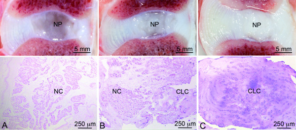
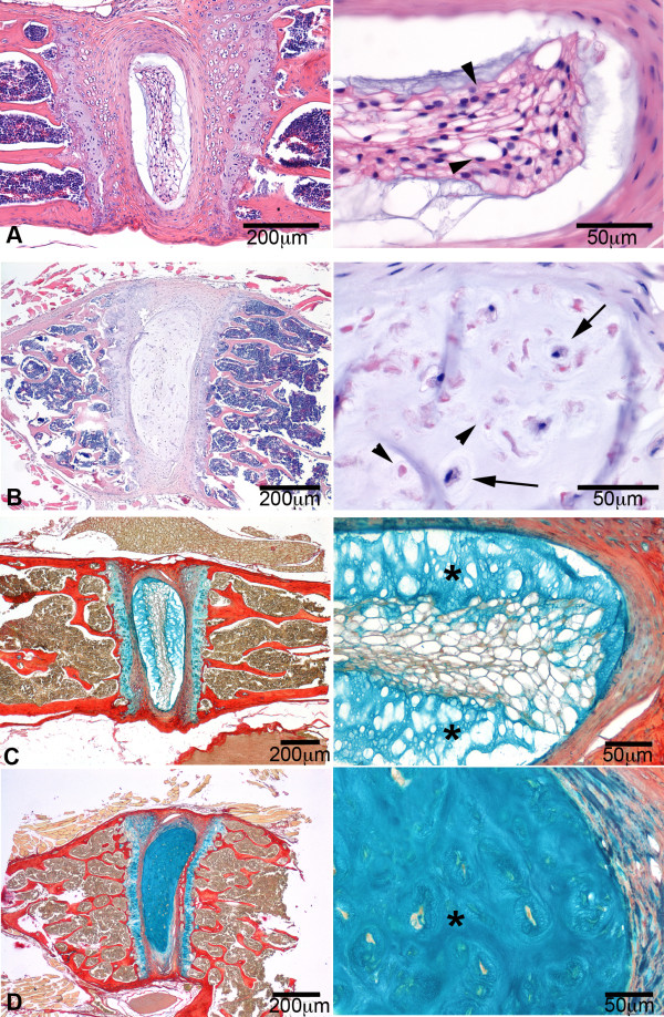
Introduction: Early degeneration of the intervertebral disc (IVD) involves a change in cellular differentiation from notochordal cells (NCs) in the nucleus pulposus (NP) to chondrocyte-like cells (CLCs). The purpose of this study was to investigate the gene expression profiles involved in this process using NP tissue from non-chondrodystrophic and chondrodystrophic dogs, a species with naturally occurring IVD degeneration.

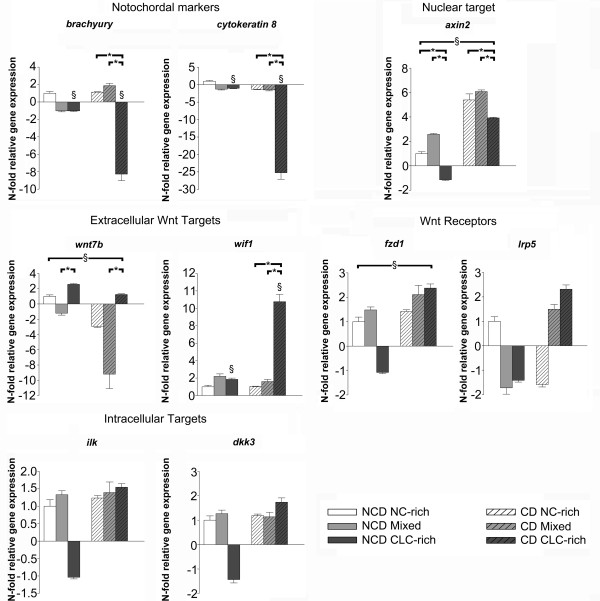
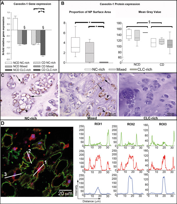
Methods: Dual channel DNA microarrays were used to compare 1) healthy NP tissue containing only NCs (NC-rich), 2) NP tissue with a mixed population of NCs and CLCs (Mixed), and 3) NP tissue containing solely CLCs (CLC-rich) in both non-chondrodystrophic and chondrodystrophic dogs. Based on previous reports and the findings of the microarray analyses, canonical Wnt signaling was further evaluated using qPCR of relevant Wnt target genes. We hypothesized that caveolin-1, a regulator of Wnt signaling that showed significant changes in gene expression in the microarray analyses, played a significant role in early IVD degeneration. Caveolin-1 expression was investigated in IVD tissue sections and in cultured NCs. To investigate the significance of Caveolin-1 in IVD health and degeneration, the NP of 3-month-old Caveolin-1 knock-out mice was histopathologically evaluated and compared with the NP of wild-type mice of the same age.

Results: Early IVD degeneration involved significant changes in numerous pathways, including Wnt/β-catenin signaling. With regard to Wnt/β-catenin signaling, axin2 gene expression was significantly higher in chondrodystrophic dogs compared with non-chondrodystrophic dogs. IVD degeneration involved significant down-regulation of axin2 gene expression. IVD degeneration involved significant down-regulation in Caveolin-1 gene and protein expression. NCs showed abundant caveolin-1 expression in vivo and in vitro, whereas CLCs did not. The NP of wild-type mice was rich in viable NCs, whereas the NP of Caveolin-1 knock-out mice contained chondroid-like matrix with mainly apoptotic, small, rounded cells.

Conclusions: Early IVD degeneration involves down-regulation of canonical Wnt signaling and Caveolin-1 expression, which appears to be essential to the physiology and preservation of NCs. Therefore, Caveolin-1 may be regarded an exciting target for developing strategies for IVD regeneration.


Figures

Figure 1
Figure 1
Typical macroscopic pictures and corresponding H&E sections of the applied classification. The notochordal cell (NC)-rich nucleus pulposus (NP) (A) contains NCs with a viable morphology organized in clusters; the mixed group (B) contains both NCs and chondrocyte-like cells (CLCs); and the CLC-rich group (C) contains solely CLCs embedded in a dense matrix.
Figure 2
Figure 2
Relative gene expression of relevant target genes. Relative gene expression of Brachyury, Cytokeratin 8, Axin2, Frizzled 1 (Fzd1), Low density lipoprotein receptor-related protein 5 (Lrp5), Wnt7b, Wnt inhibitory factor 1 (Wif1), Integrin linked kinase (Ilk), and Dickkopf homolog 3 (Dkk3) in the notochordal cell-rich (NC-rich), mixed, and chondrocyte-like cell rich (CLC-rich) NP from non-chondrodystrophic (NCD) and chondrodystrophic (CD) dogs (NCD, NC-rich NP was used as reference, set at 1). *Significant difference between NC-rich, mixed, and CLC-rich NP; §significant difference between NCD and CD dogs.
Figure 3
Figure 3
Caveolin-1 expression in the course of early intervertebral disc degeneration. (A) Caveolin-1 gene expression and (B) proportion of the NP surface area that stained for Caveolin-1, and mean gray value for Caveolin-1 protein expression in the notochordal cell (NC)-rich, mixed, and chondrocyte-like cell (CLC)-rich nucleus pulposus (NP) from non-chondrodystrophic (NCD) and chondrodystrophic (CD) dogs. *Significant difference between degeneration stages; §significant difference between NCD and CD dogs. The proportion of the NP surface area that stained for caveolin-1 was not divided into NCD and CD dogs because no significant differences were found between breed types. (C) Typical examples of NP samples stained for Caveolin-1, showing the NC-rich NP, mixed cell population NP with NCs and CLCs, and the CLC-rich NP. In the NC-rich and mixed groups, membranous (arrow) and cytoplasmic (arrowhead) staining can be observed. Note that Caveolin-1 staining is not observed in CLCs. (D) Immunofluorescent staining of the NC-rich NP for the proteins Caveolin-1 (green) and β-catenin (red), and for DNA (blue). Region of interest (ROI) lines drawn across cell bodies were used to generate profile intensity plots (right) for the signal intensity of the Caveolin-1 (green), β-catenin (red), and Topro-3 (blue). The signal intensity peaks for Caveolin-1 correspond with the signal peaks of β-catenin at the cell membrane (located at the same distance of the ROI line), indicating colocalization of these proteins. Also, the central β-catenin signal peaks correspond with the Topro-3 signal peaks, indicating nuclear localization of β-catenin.
Figure 4
Figure 4
Immunofluorescence of Caveolin-1 in primary notochordal cells in monolayer culture. Immunofluorescence images of the notochordal cell clusters on days 0, 2, 4, 6, 8, and 10 in culture. Scale bar: 50 μm. Nuclear staining (Topro-3) and Caveolin-1 staining are depicted in blue and green, respectively. Caveolin-1 protein was located in intracellular membranes, as suggested by the inhomogeneous appearance of the immunolabeled membrane-embedded marker, and in the notochordal cell membrane.
Figure 5
Figure 5
The intervertebral disc phenotype of Caveolin-1 knock-out mice. Typical examples of the intervertebral discs from 3-month-old wild-type (A and C) and Caveolin-1 knock-out (B and D) mice, stained with H&E (A and B) and alcian blue/pricrosirius red (C and D). The pictures on the right are magnifications of the corresponding pictures on the left. The wild-type nucleus pulposus (NP) consisted of centrally located, viable notochordal cells (arrowhead) and a relatively limited amount of chondroid-like intercellular matrix (*), which stains blue in the sections stained with alcian blue/picrosirius red. The NP of the Caveolin-1 KO mice contained apoptotic (arrowheads) and rounded cells, with a smaller amount of cytoplasm lacking the typical vacuolar aspect (arrows), and a relatively large amount of chondroid-like intercellular matrix (*).

References

    1. Luoma K, Riihimaki H, Luukkonen R, Raininko R, Viikari-Juntura E, Lamminen A. Low back pain in relation to lumbar disc degeneration. Spine (Phila Pa 1976) 2000;25:487–492. doi: 10.1097/00007632-200002150-00016. - DOI - PubMed
    1. Bono CM, Lee CK. Critical analysis of trends in fusion for degenerative disc disease over the past 20 years: influence of technique on fusion rate and clinical outcome. Spine (Phila Pa 1976) 2004;29:455–463. doi: 10.1097/01.BRS.0000090825.94611.28. discussion Z455. - DOI - PubMed
    1. Gibson JN, Waddell G. Surgical interventions for lumbar disc prolapse: updated Cochrane Review. Spine (Phila Pa 1976) 2007;32:1735–1747. doi: 10.1097/BRS.0b013e3180bc2431. - DOI - PubMed
    1. Brinckmann P, Grootenboer H. Change of disc height, radial disc bulge, and intradiscal pressure from discectomy. An in vitro investigation on human lumbar discs. Spine (Phila Pa 1976) 1991;16:641–646. doi: 10.1097/00007632-199106000-00008. - DOI - PubMed
    1. Eck JC, Humphreys SC, Hodges SD. Adjacent-segment degeneration after lumbar fusion: a review of clinical, biomechanical, and radiologic studies. Am J Orthop. 1999;28:336–340. - PubMed

Publication types

MeSH terms