Review
. 2013 May;104(5):525-30.
doi: 10.1111/cas.12118. Epub 2013 Mar 11.

Roles and mechanisms of cellular senescence in regulation of tissue homeostasis

Affiliations
Review

Roles and mechanisms of cellular senescence in regulation of tissue homeostasis

Naoko Ohtani et al. Cancer Sci. 2013 May.

Abstract

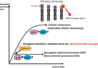
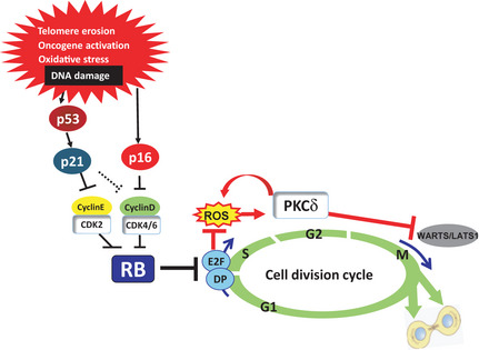
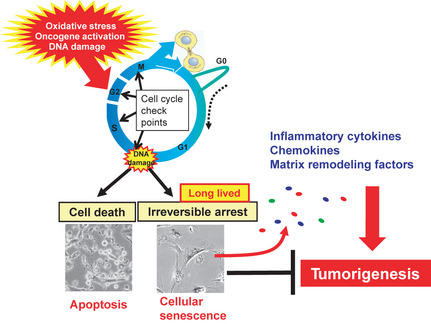
Cellular senescence is the state of irreversible cell cycle arrest that can be induced by a variety of potentially oncogenic stimuli and has therefore long been considered to suppress tumorigenesis, acting as a guardian of homeostasis. However, surprisingly, emerging evidence reveals that senescent cells also promote secretion of a series of inflammatory cytokines, chemokines, growth factors and matrix remodeling factors, which alter the local tissue environment and contribute to chronic inflammation and cancer. This newly identified senescence phenotype, termed the senescence-associated secretory phenotype (SASP) or the senescence-messaging secretome (SMS), is induced by DNA damage that promotes the induction of cellular senescence. All of these senescence-associated secreting factors are involved in homeostatic disorders such as cancer. Therefore, it is quite possible that accumulation of senescent cells during the aging process in vivo might contribute to age-related increases in homeostatic disorders. In this review, current knowledge of the molecular and cellular biology of cellular senescence is introduced, focusing on its positive and negative roles in controlling tissue homeostasis in vivo.


Figures

Figure 1
Figure 1
What is cellular senescence? Most normal human somatic cells stop dividing after a finite number of cell divisions and enter a state of irreversible cell proliferation arrest. This phenomenon is called “cellular senescence” or “replicative cellular senescence”. In human cells, the mechanism underlying replicative cellular senescence is thought to be telomere shortening. Recent studies in human cells revealed that a similar irreversible proliferation block can be induced quite rapidly when normal cells are exposed to a variety of potentially oncogenic stimuli, such as excessive levels of reactive oxygen species, treatment with DNA damaging agents or activation of certain oncogenes. Because all of these stimuli induce irreparable DNA damage in both human and murine cells, it is plausible that persistent DNA damage signals induce cellular senescence. These types of oncogenic stress‐induced senescence are now referred to as “oncogene‐induced senescence” or “stress‐induced senescence”. Both p16 and p21 upregulation contributes to the induction of both types of cellular senescence.
Figure 2
Figure 2
Molecular mechanisms of cellular senescence. A series of cyclin‐dependent kinases (CDK) including CDK2, CDK4 and CDK6 play key roles in regulating the activities of RB‐family proteins. When RB is phosphorylated by these CDK, it loses its ability to bind to and repress the functions of the E2F family of transcription factors, thereby resulting in gene transcription required for the initiation of DNA replication and cell cycle progression. In response to irreparable DNA damage caused by a variety of oncogenic stimuli, expression levels of the p21 Cip1 and p16 Ink4a CDK inhibitors are significantly upregulated by p53‐dependent and p53‐independent mechanisms, respectively. Activation of the p16Ink4a–Rb pathway cooperates with mitogenic signals to induce elevated intracellular levels of reactive oxygen species (ROS), thereby causing activation of PKCδ, a critical downstream mediator of the ROS signaling pathway, and leading to a cytokinetic block that might provide an additional safeguard against the proliferation of senescent cells, especially in the case of RB and p53 subsequently become inactivated.
Figure 3
Figure 3
Dynamics of p16 Ink4a gene expression during the aging process in vivo. The p16‐luc mice, an imaging mice for p16 gene expression in vivo, were subjected to non‐invasive bioluminescence imaging throughout their entire life span. Accumulation of p16 was confirmed in the aged mouse.
Figure 4
Figure 4
Senescence‐associated secretome. Irreparable DNA damage provokes either apoptosis or cellular senescence depending on the strength of stress and/or cellular context. Recently it has become apparent that long‐lived senescent cells exhibit increased expression of genes encoding a series of secreted proteins, such as inflammatory cytokines, chemokines and matrix remodeling factors, which alter the local tissue environment and/or contribute to chronic inflammation and tumorigenesis. Cellular senescence initially prevents proliferation of damaged cells, thereby acting as a fail‐safe mechanism. However, in the long term, senescent cells might eventually promote tumorigenesis by accelerating chromosomal instability and/or production of senescence‐associated secretome factors.

Similar articles

Cited by

References

    1. Martin GM. The biology of aging: 1985–2010 and beyond. FASEB J 2011; 25: 3756–62. - PMC - PubMed
    1. Finkel T, Serrano M, Blasco MA. The common biology of cancer and ageing. Nature 2007; 448: 767–74. - PubMed
    1. Campisi J. Senescent cells, tumor suppression, and organismal aging: good citizens, bad neighbors. Cell 2005; 120: 513–22. - PubMed
    1. Herbig U, Sedivy JM. Regulation of growth arrest in senescence: telomere damage is not the end of the story. Mech Ageing Dev 2006; 127: 16–24. - PubMed
    1. Sharpless NE, DePinho RA. Cancer: crime and punishment. Nature 2005; 436: 636–7. - PubMed

Publication types

MeSH terms