Case Reports
. 2013 Jan 29:8:22.
doi: 10.1186/1749-8090-8-22.

Surgical excision with left atrial reconstruction of a primary functioning retrocardiac paraganglioma

Affiliations
Case Reports

Surgical excision with left atrial reconstruction of a primary functioning retrocardiac paraganglioma

María Teresa González López et al. J Cardiothorac Surg. .

Abstract

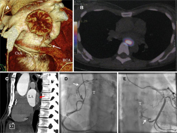
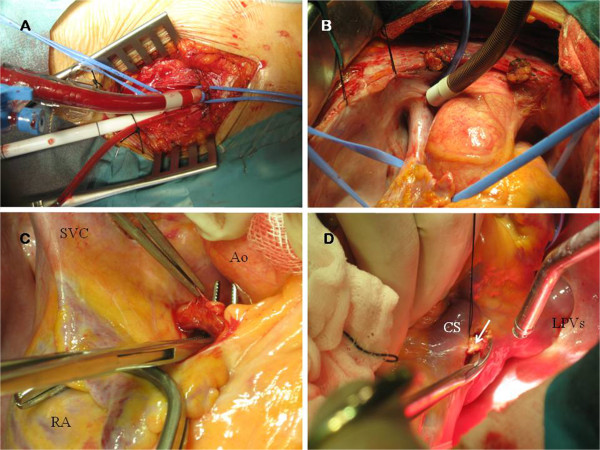
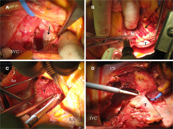
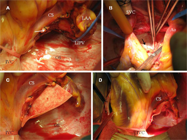
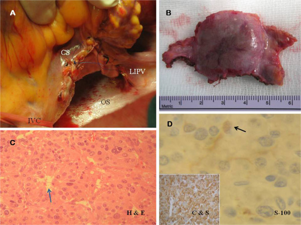
About 2% of all paragangliomas are located in the chest, and a few have been described to be found in the heart. Primary cardiac paragangliomas are extremely uncommon tumors and surgical experience with this neoplasm is limited. Treatment strategies described in the literature have included simple excision, excision with reconstruction, autotransplantation after excision of the tumor and even orthotopic cardiac transplantation, depending on the extent of disease. A primary retrocardiac paraganglioma catecholamine-productive was identified in an asymptomatic 49-year old female associated to familial pheochromocytoma-paraganglioma syndrome caused by germline mutation of the gen which codifies for the subunit B of succinate dehydrogenase enzyme (SDHB). The neoplasm was surgically excised from the posterior surface of the left atrium via median sternotomy using cardiopulmonary bypass. Direct ligation of feeding vessels of the tumor along with left atrial reinforcement using a pericardial patch was performed. The post-operative course was uneventful, with normalization of catecholamine secretion and no recurrence at three-month follow-up. We review the current literature about this exceptional cardiac tumor, pathophysiological conditions and options for surgical management.


Figures

Figure 1
Figure 1
A. Computed tomography imaging reconstruction of the heart. The tumor is showed. The collateral vessel (white arrow) from the circumflex artery (CxA) is observed. B. 18-F DOPA PET showed a 16 × 25 × 36 mm extracardiac mass protruding within the LA. Presence of metabolic activity was detected. C. Computed tomography (axial view) showed the presence of the large mass (red arrow). D. Coronary angiography. The coronary blood supply of the superior aspect of the paraganglioma (T) emerged from a large collateral vessel (white arrow) from the right coronary artery (RCA). E. Coronary angiography. Proximal and distal collaterals from CxA were the feeding vessels (white arrow) of the inferior aspect of the tumor (T).
Figure 2
Figure 2
A. Peripheral perfusion through right femoral artery and vein. B. Superior vein cavae (SVC) drainage through Pacifico cannula.C. The large collateral vessel from the RCA was dissected and ligated. RA: Right atrium. Ao: Aorta. D. The two collateral vessels from CxA (white arrow) were also ligated for an optimal control of bleeding during the excision of the tumor. CS: Coronary sinus. LPVs: Left pulmonary veins.
Figure 3
Figure 3
A. Surgeon’s view. Superior aspect of the tumor (black arrow) is showed. Neovascularization of the smooth surface is observed. B. Endocardium of the LA was exposed (black arrow) during surgical excision (suspended heart). C. Surgeon’s view. Dissection of the superior aspect of the tumor (T). D. Paraganglioma (T) after totally excision from the LA. IVC: Inferior vein cava. LIPV: Left inferior vein cava.
Figure 4
Figure 4
A. LA after removal of the tumor. The operative photograph shows the distal collateral vessel (white arrow) from CxA (after ligation). Endocardium of the LA remained in situ. LAA: Left atrial appendage. OS: Oblique sinus. B. Bovine pericardial patch (black arrow) was used for left atrial reconstruction. The suture was started from the left atrial roof. C-D. Pericardial patch was placed on the posterior wall of the LA using a polypropylene running suture.
Figure 5
Figure 5
A. Posterior LA after pericardial replacement. B. Macroscopic examination (6 × 3 × 1.5 cm fragment). A well-demarcated homogeneous, smooth surfaced and highly vascularized tumor (2.8 × 2.5 × 1.5 cm) was found, with clear surgical margins. C. Histological examination. Haematoxylin and eosin staining (H & E, 40x magnification) showing the “zellballen” packeting of cells, without atypia, surrounded by capillary network (blue arrow). D. Immunohistochemical analysis (40x magnification) revealed the specific neuroendocrine markers (chromogranin and synaptophysin, C & S). S-100 protein staining technique (100x magnification) revealed the sustentacular cell network (black arrow).

References

    1. Walther MM, Keiser HR, Linehan WM. Pheochromocytoma: evaluation, diagnosis, and treatment. World J Urol. 1999;17:35–39. doi: 10.1007/s003450050102. - DOI - PubMed
    1. Manger WM. In search of pheochromocytomas. J Clin Endocrinol Metab. 2003;88:4080–4082. doi: 10.1210/jc.2003-031234. - DOI - PubMed
    1. Pacheco N, Marcos G, Garcipérez FJ, Pérez C. Intrapericardial paraganglioma. Rev Esp Cardiol. 2010;63:116–117. - PubMed
    1. Elder EE, Elder G, Larsson C. Pheochromocytoma and functional paraganglioma syndrome: no longer the 10% tumor. J Surg Oncol. 2005;89:193–201. doi: 10.1002/jso.20177. - DOI - PubMed
    1. Tomasian A, Lai C, Ruehm S, Krishnam MS. Cardiovascular magnetic resonance and PET-CT of left atrial paraganglioma. J Cardiovasc Magn Reson. 2010;12:1. doi: 10.1186/1532-429X-12-1. - DOI - PMC - PubMed

Publication types