. 2013 Jan 29:8:17.
doi: 10.1186/1750-1172-8-17.

Passive transfer of collagen XVII-specific antibodies induces sustained blistering disease in adult mice

Affiliations

Passive transfer of collagen XVII-specific antibodies induces sustained blistering disease in adult mice

Mircea Teodor Chiriac et al. Orphanet J Rare Dis. .

Abstract

Background: Bullous pemphigoid is a subepidermal blistering disorder associated with tissue-bound and circulating autoantibodies directed mainly to the hemidesmosomal component collagen XVII. While recapitulating the main immunopathological features of the human disease, frank skin blistering does not develop in the absence of skin rubbing in experimental pemphigoid models that have been established in neonatal mice. Moreover, due to their experimental design they only allow for short-term disease observation. In the present study we aimed to establish a model that reproduces the frank skin blistering seen in patients and allows for longer observation times.

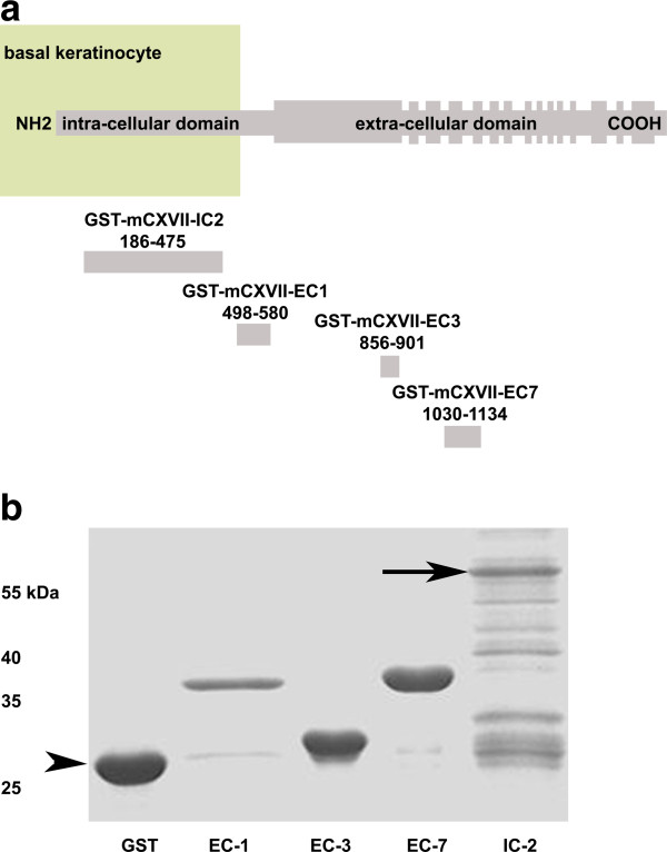
Methods: Rabbit and sheep antibodies specific to several fragments of collagen XVII were generated and the purified antibodies were passively transferred into adult mice.

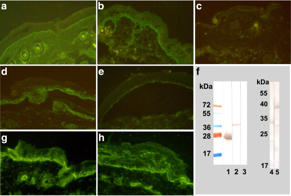
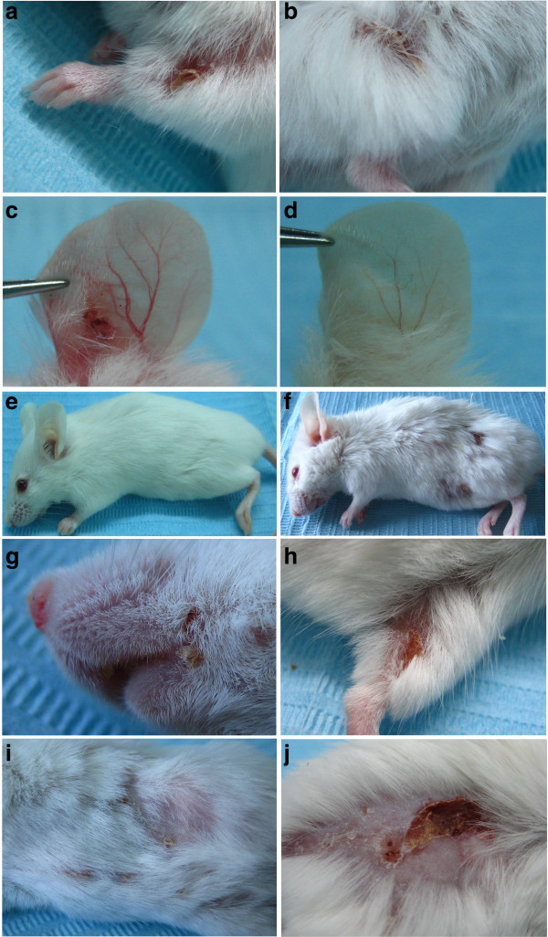
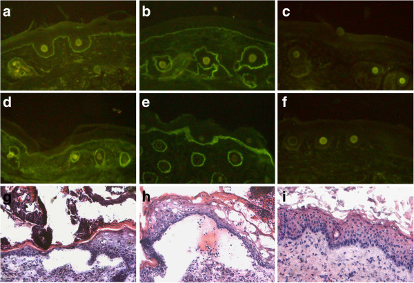
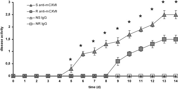
Results: Collagen XVII-specific IgG bound to the basal membrane of the skin and mucous membranes activating murine complement in vivo. Mice injected with collagen XVII-specific antibodies, in contrast to mice receiving control antibodies, developed frank skin blistering disease, reproducing human bullous pemphigoid at the clinical, histological and immunopathological levels. Titres of circulating IgG in the serum of mice correlated with the extent of the clinical disease. Mice receiving sheep antibodies specific to murine collagen XVII showed an early onset and a more active disease when compared to litter mates receiving specific rabbit antibodies.

Conclusion: This novel animal model for bullous pemphigoid should facilitate further investigations of the pathogenesis of bullous pemphigoid and the development of innovative therapies for this disease.


Figures

Figure 1
Figure 1
Recombinant fragments of murine BP180/ CXVII. (a) BP180/ CXVII is composed of an intracellular N-terminal fragment localized to the hemidesmosomal plaque of basal keratinocytes with a long extracellular portion containing 15 collagenous domains (filled rectangles) separated by non-collagenous regions protruding into the basal membrane. Four fragments of murine type XVII collagen cDNA cloned in pGEX-6P-1 were expressed in E. coli. Amino acid positions are shown below each fragment (the diagram is not at scale). (b) When run on an SDS-PAGE gel the proteins migrated correspondingly to their calculated molecular weights of 37, 32, 39 and 57 kDa, respectively. In lanes 1 through 6 we loaded: molecular weight marker, GST, GST-mCXVII-EC1, GST-mCXVII-EC3, GST-mCXVII-EC7 and GST-mCXVII-IC2. The arrow indicates GST-mCXVII-IC2 at 57kDa and the arrowhead indicates GST at 27kDa.
Figure 2
Figure 2
Antibodies from immunized animals recognize mouse BP180/ CXVII and activate complementex vivo. Serum was obtained from immunized animals at different time points. Immune rabbit (a) and sheep (b) antibodies bound to the basal membrane of the mouse skin. In contrast, antibodies obtained from a rabbit before the first immunization (c) did not recognize the antigen in situ. When incubated with mouse split skin, IgG from the immune sera of both the rabbit (d) and sheep (e) bound to the epidermal side of the 1 NaCl-split skin (magnifications 200x). (f) Pathogenic antibodies in contrast to control antibodies recognized recombinant forms of BP180/ CXVII by immunoblotting; in the left hand panel: 1, GST and 2, GST-mCXVII-EC1 incubated with immune rabbit serum; 3, GST-mCXVII-EC1 incubated with control rabbit antibodies as a negative control; in the right hand panel: 4, GST-mCXVII-EC3 and 5, GST-mCXVII-EC7 incubated with immune rabbit serum. BP180/ CXVII-specific antibodies from the rabbit (g) and sheep (h) activated complement when incubated on cryosections of murine skin (magnifications 200x).
Figure 3
Figure 3
BP180/ CXVII-specific antibodies induce blistering skin disease when passively transferred into adult mice. Mice receiving rabbit antibodies (n=10) to murine BP180/ CXVII developed skin lesions at different sites including the (a) front limbs, the (b) hind limbs and the (c) ears. In contrast, the ear of a mouse receiving control rabbit antibodies remained unaffected (d) as did a mouse receiving control sheep antibodies (e). Mice injected with (f) pathogenic sheep antibodies (n=10) presented an even more extensive and generalized disease. Lesions including erosions partly covered by crusts were recorded on the (g) snout, and (h) hind limb of a mouse receiving sheep antibodies to murine BP180/ CXVII. More advanced disease was characterized by blisters, erosions and alopecia affecting both the (i) abdomen and the (j) back of mice injected with sheep BP180/ CXVII-specific IgG.
Figure 4
Figure 4
Diseased mice show immuno- and histo-pathological features of the pemphigoid disease. At the end of the observation period, mice were sacrificed and perilesional biopsies were analyzed for immunoreactants deposition. By IF microscopy, the perilesional skin of a diseased mouse injected with (a) rabbit or (b) sheep pathogenic antibodies showed linear deposition of murine IgG at the DEJ. Additionally, tissue bound (d) rabbit and (e) sheep antibodies were shown to activate murine C3 in vivo as revealed by direct IF microscopy. In contrast, no (c) IgG or (f) complement deposits were found in mice injected with control sheep antibodies. The lesional skin of a mouse receiving (g) rabbit or (h) sheep BP180/ CXVII-specific IgG shows a dermal-epidermal separation accompanied by an inflammatory infiltrate. In contrast, the skin of a mouse receiving (i) control sheep antibodies showed no histopathological signs of disease (magnifications, 200x).
Figure 5
Figure 5
Anti-murine type XVII collagen antibodies of sheep precipitate the onset and potentiate the outcome of disease. Mice were injected with antibodies to type XVII collagen generated in rabbits (n=10) and sheep (n=10) respectively. After 9–10 days, first lesions appeared in mice transferred with antibodies produced in rabbits. In contrast, mice receiving antibodies generated in sheep presented first lesions after 5–6 days. In addition, at each time-point of the observation period, starting with the day of first lesions appearance, the group injected with pathogenic sheep antibodies had significantly higher disease scores compared to the group injected with pathogenic rabbit anti-mouse collagen BP180/ CXVII antibodies. Groups injected with either control rabbit (n=6) or sheep (n=6) antibodies did not show any clinical lesions throughout the observation period. Means are presented ± s.e.m.; *, significance at p < 0.05 by the chi-square test.

Similar articles

Cited by

References

    1. Zillikens D, Wever S, Roth A, Weidenthaler-Barth B, Hashimoto T, Bröcker EB. Incidence of autoimmune subepidermal blistering dermatoses in a region of central Germany. Arch Dermatol. 1995;131:957–958. doi: 10.1001/archderm.1995.01690200097021. - DOI - PubMed
    1. Bernard P, Vaillant L, Labeille B, Bedane C, Arbeille B, Denoeux JP, Lorette G, Bonnetblanc JM, Prost C. Incidence and distribution of subepidermal autoimmune bullous skin diseases in three french regions. Bullous diseases french study group. Arch Dermatol. 1995;131:48–52. doi: 10.1001/archderm.1995.01690130050009. - DOI - PubMed
    1. Langan SM, Smeeth L, Hubbard R, Fleming KM, Smith CJ, West J. Bullous pemphigoid and pemphigus vulgaris--incidence and mortality in the UK: population based cohort study. BMJ. 2008;337:a180. doi: 10.1136/bmj.a180. - DOI - PMC - PubMed
    1. Marazza G, Pham HC, Scharer L, Pedrazzetti PP, Hunziker T, Trueb RM, Hohl D. et al.Incidence of bullous pemphigoid and pemphigus in Switzerland: a 2-year prospective study. Br J Dermatol. 2009;161:861–868. doi: 10.1111/j.1365-2133.2009.09300.x. - DOI - PubMed
    1. Stanley JR, Hawley-Nelson PYuspa SH, Shevach EM, Katz SI. Characterization of bullous pemphigoid antigen: a unique basement membrane protein of stratified squamous epithelia. Cell. 1981;24:897–903. doi: 10.1016/0092-8674(81)90115-X. - DOI - PubMed

Publication types

LinkOut - more resources