Intravenous contrast material-induced nephropathy: causal or coincident phenomenon?
- PMID: 23360742
- PMCID: PMC6940002
- DOI: 10.1148/radiol.12121823
Intravenous contrast material-induced nephropathy: causal or coincident phenomenon?
Erratum in
-
Intravenous Contrast Material-induced Nephropathy: Causal or Coincident Phenomenon?Radiology. 2016 Jan;278(1):306. doi: 10.1148/radiol.2015154044. Radiology. 2016. PMID: 26690999 Free PMC article. No abstract available.
Abstract
Purpose: To determine the causal association and effect of intravenous iodinated contrast material exposure on the incidence of acute kidney injury (AKI), also known as contrast material-induced nephropathy (CIN).
Materials and methods: This retrospective study was approved by an institutional review board and was HIPAA compliant. Informed consent was waived. All contrast material-enhanced (contrast group) and unenhanced (noncontrast group) abdominal, pelvic, and thoracic CT scans from 2000 to 2010 were identified at a single facility. Scan recipients were sorted into low- (<1.5 mg/dL), medium- (1.5-2.0 mg/dL), and high-risk (>2.0 mg/dL) subgroups of presumed risk for CIN by using baseline serum creatinine (SCr) level. The incidence of AKI (SCr ≥ 0.5 mg/dL above baseline) was compared between contrast and noncontrast groups after propensity score adjustment by stratification, 1:1 matching, inverse weighting, and weighting by the odds methods to reduce intergroup selection bias. Counterfactual analysis was used to evaluate the causal relation between contrast material exposure and AKI by evaluating patients who underwent contrast-enhanced and unenhanced CT scans during the study period with the McNemar test.
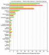
Results: A total of 157,140 scans among 53,439 unique patients associated with 1,510,001 SCr values were identified. AKI risk was not significantly different between contrast and noncontrast groups in any risk subgroup after propensity score adjustment by using reported risk factors of CIN (low risk: odds ratio [OR], 0.93; 95% confidence interval [CI]: 0.76, 1.13; P = .47; medium risk: odds ratio, 0.97; 95% CI: 0.81, 1.16; P = .76; high risk: OR, 0.91; 95% CI: 0.66, 1.24; P = .58). Counterfactual analysis revealed no significant difference in AKI incidence between enhanced and unenhanced CT scans in the same patient (McNemar test: χ(2) = 0.63, P = .43) (OR = 0.92; 95% CI: 0.75, 1.13; P = .46).
Conclusion: Following adjustment for presumed risk factors, the incidence of CIN was not significantly different from contrast material-independent AKI. These two phenomena were clinically indistinguishable with established SCr-defined criteria, suggesting that intravenous iodinated contrast media may not be the causative agent in diminished renal function after contrast material administration.
Supplemental material: http://radiology.rsna.org/lookup/suppl/doi:10.1148/radiol.12121823/-/DC1.
RSNA, 2013
Figures


Comment in
-
Quantitating contrast medium-induced nephropathy: controlling the controls.Radiology. 2013 Apr;267(1):4-8. doi: 10.1148/radiol.13122876. Radiology. 2013. PMID: 23525714 No abstract available.
References
-
- Barrett BJ, Parfrey PS. Clinical practice: preventing nephropathy induced by contrast medium. N Engl J Med 2006;354(4):379–386. - PubMed
-
- Gleeson TG, Bulugahapitiya S. Contrast-induced nephropathy. AJR Am J Roentgenol 2004;183(6):1673–1689. - PubMed
-
- Solomon R, Dumouchel W. Contrast media and nephropathy: findings from systematic analysis and Food and Drug Administration reports of adverse effects. Invest Radiol 2006;41(8):651–660. - PubMed
-
- Tepel M, Aspelin P, Lameire N. Contrast-induced nephropathy: a clinical and evidence-based approach. Circulation 2006;113(14):1799–1806. - PubMed
-
- Tublin ME, Murphy ME, Tessler FN. Current concepts in contrast media-induced nephropathy. AJR Am J Roentgenol 1998;171(4):933–939. - PubMed
MeSH terms
Substances
Grants and funding
LinkOut - more resources
Full Text Sources
Other Literature Sources
Medical

