. 2013 Sep;6(5):1027-37.
doi: 10.1038/mi.2012.141. Epub 2013 Jan 30.

The functional maturation of M cells is dramatically reduced in the Peyer's patches of aged mice

Affiliations
Free PMC article

The functional maturation of M cells is dramatically reduced in the Peyer's patches of aged mice

A Kobayashi et al. Mucosal Immunol. 2013 Sep.
Free PMC article

Abstract

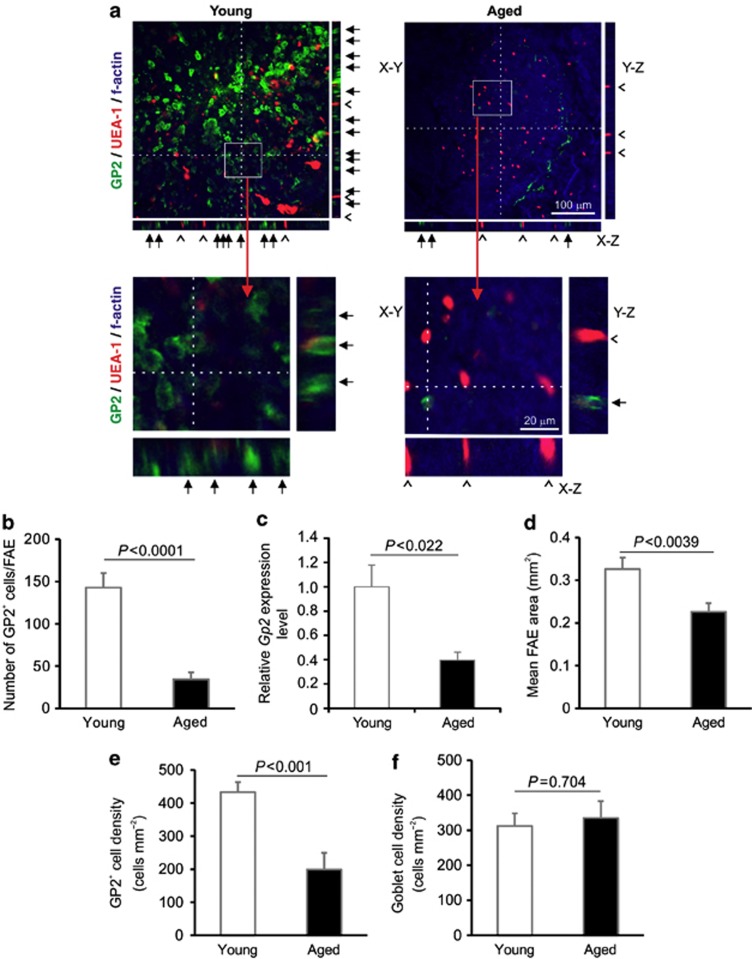
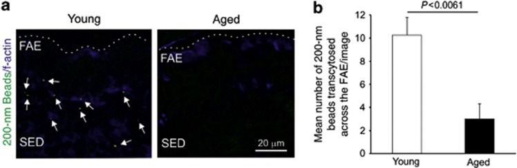
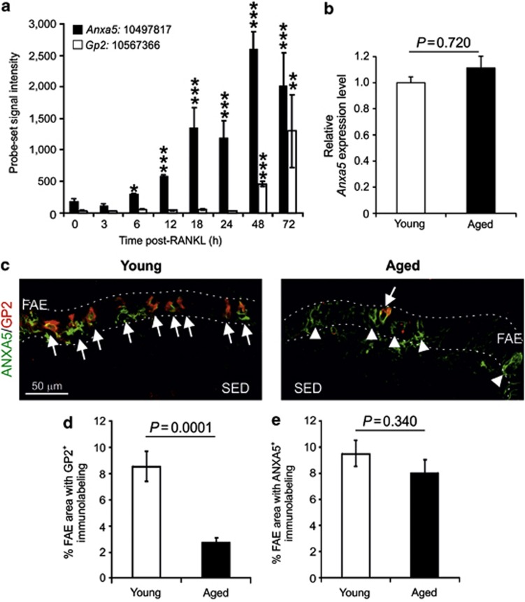
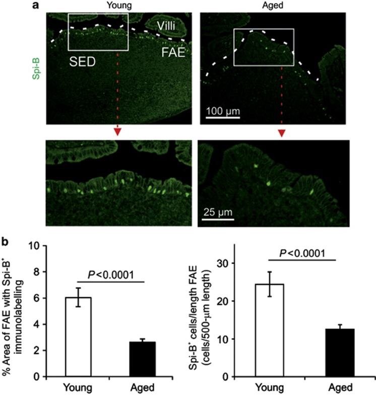
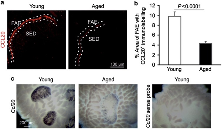
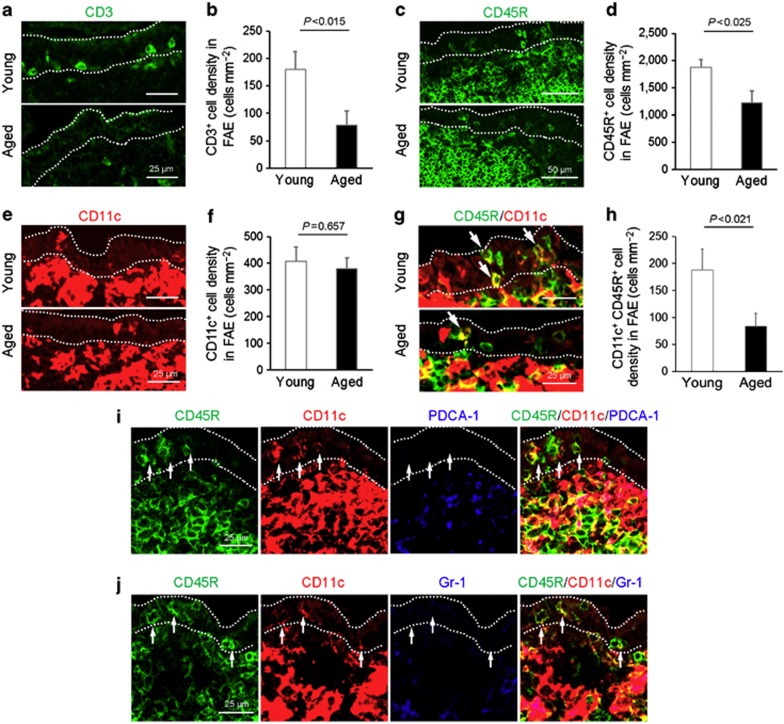
The transcytosis of antigens across the follicle-associated epithelium (FAE) of Peyer's patches by microfold cells (M cells) is important for the induction of efficient immune responses to mucosal antigens. The mucosal immune response is compromised by ageing, but effects on M cells were unknown. We show that M-cell density in the FAE of aged mice was dramatically reduced. As a consequence, aged Peyer's patches were significantly deficient in their ability to transcytose particulate lumenal antigen across the FAE. Ageing specifically impaired the expression of Spi-B and the downstream functional maturation of M cells. Ageing also dramatically impaired C-C motif chemokine ligand 20 expression by the FAE. As a consequence, fewer B cells were attracted towards the FAE, potentially reducing their ability to promote M-cell maturation. Our study demonstrates that ageing dramatically impedes the functional maturation of M cells, revealing an important ageing-related defect in the mucosal immune system's ability to sample lumenal antigens.


Figures

Figure 1
Figure 1
Microfold cell (M-cell) density is significantly reduced in the follicle-associated epithelium (FAE) of Peyer's patches from aged mice. (a) Tissues were immunostained to detect glycoprotein 2 (GP2; green), Ulex europaeus agglutinin-1 (UEA-1; red) and f-actin (blue). The positions of the X-Z and Y-Z projections of the FAE are indicated by the broken line in the main X-Y images. Closed arrows indicate GP2+ M cells with characteristic basolateral pockets. Open arrowheads indicate GP2-UEA-1+ goblet cells. The boxed areas in each of the main X-Y images are shown below at higher magnification. (b) Morphometric analysis indicated that the number of GP2+ M cells in the FAE of aged mice was significantly reduced (P<0.0001, Mann–Whitney U test). (c) Quantitative real-time reverse transcriptase–PCR analysis revealed a significant reduction in Gp2 transcript levels in tissues from aged mice (P<0.022, Student's t-test). Gene expression data are normalized so that the mean level in young mice was 1.0. (d) Morphometric analysis suggested that the size of the FAE in Peyer's patches of aged mice was smaller than those in young mice (P<0.039, Student's t-test). (e) The density of M cells in the FAE of aged mice was significantly reduced (P<0.001, Mann–Whitney U test). (f) Ageing did not influence the density of GP2-UEA-1+ goblet cells in the FAE (P=0.707, Student's t-test). Data are derived from 3–5 Peyer's patches from four mice from each group.
Figure 2
Figure 2
The uptake of particulate antigen into the Peyer's patches of aged mice is significantly impaired. (a) In Peyer's patches from young mice, many fluorescent beads (arrows, green) had been transcytosed across the follicle-associated epithelium (FAE). Substantially fewer, if any, beads appeared to have passed through the FAE of aged mice. Sections were counterstained to detect f-actin (blue). Broken line indicates the lumenal surface of the FAE. (b) The number of beads transcytosed across the FAE of aged mice was significantly less than that observed in Peyer's patches from young mice (P<0.0061, Student's t-test). Data were collected from 10 sections from each Peyer's patch. Data are derived from 3–5 Peyer's patches from four mice from each group. SED, subepithelial dome.
Figure 3
Figure 3
Ageing does not influence the expression of receptor activator of NF-κB ligand (RANKL), RANK, and osteoprotegerin (OPG) in Peyer's patches. (a) Immunohistochemical (IHC) analysis suggested that there was no observable difference in the expression or distribution of RANKL on subepithelial dome (SED) of the stromal cells in Peyer's patches from young and aged mice. Broken line indicates the lumenal surface of the follicle-associated epithelium (FAE). (b) Morphometric analysis confirmed that the magnitude of RANKL-specific immunostaining observed in the SED of Peyer's patches from young and aged mice was similar. Quantitative real-time reverse transcriptase–PCR analysis suggested that there was no significant difference in the expression of (c) Tnfsf11 (RANKL), (d) Tnfrsf11a (RANK), or (e) Tnfrsf11b (OPG) mRNA levels between Peyer's patches from young or aged mice. Gene expression data are normalized so that the mean level in young mice was 1.0. Data are derived from 3–5 Peyer's patches from at least four mice/group.
Figure 4
Figure 4
Ageing does not affect the expression of annexin A5 (ANXA5) in the follicle-associated epithelium (FAE). (a) Effect of systemic recombinant-RANKL (receptor activator of NF-κB ligand) treatment on the expression of Anxa5 (closed bars) and glycoprotein 2 (Gp2; open bars) in the villous epithelium. Each bar represents mean probe-set signal intensity. The notation XXXX:12345678 represents gene symbol:Affymetrix probe-set ID. *P<0.05; **P<0.002; P<0.0001 (Student's t-test). (b) Quantitative real-time reverse transcriptase–PCR analysis suggested that there was no significant difference in the expression levels of Anxa5 in young or aged mice. (c) Immunohistochemical analysis of the expression of GP2 (red) and ANXA5 (green) by M cells in the FAE of young and aged mice. Arrows, GP2+ANXA5+ M cells; arrowheads, GP2-ANXA5+ immature M cells. Broken lines indicate the boundary of the FAE. (d) Morphometric analysis confirmed that the magnitude of the GP2-specific immunostaining observed in the FAE of Peyer's patches from aged mice was significantly reduced when compared with young mice (P<0.0001, Mann–Whitney U test). (e) However, ageing did not influence the magnitude of the ANXA5-specific immunostaining observed in the FAE (P=0.340, Student's t-test). Data are derived from 3–5 Peyer's patches from at least four mice from each group. SED, subepithelial dome.
Figure 5
Figure 5
Effects of ageing on Spi-B expression in the follicle-associated epithelium (FAE) of Peyer's patches. (a) Immunohistochemical analysis suggested that Spi-B (green) was reduced in the FAE of aged mice. Boxed area in upper panels is shown at higher magnification in lower panels. Broken line indicates the lumenal surface of the FAE. (b) Morphometric analysis showed that the magnitude of the Spi-B-specific immunostaining and number of Spi-B+ cells were significantly reduced in the FAE of aged mice (P<0.0001, Student's t-test). Data are derived from 4–6 Peyer's patches from eight mice from each group. SED, subepithelial dome.
Figure 6
Figure 6
Effects of ageing on C-C motif chemokine ligand 20 (CCL20) expression in the follicle-associated epithelium (FAE) of Peyer's patches. (a) Immunohistochemical analysis suggested that CCL20 (red) was reduced in the FAE of aged mice. Broken line indicates the lumenal surface of the FAE. (b) Morphometric analysis showed that the magnitude of CCL20-specific immunostaining observed in aged mice was significantly reduced (P<0.0001, Student's t-test). Data are representative of 3–5 Peyer's patches from eight mice from each group. (c) Whole-mount in situ hybridization analysis demonstrated a dramatic reduction in Ccl20 mRNA expression in the Peyer's patches of aged mice when compared with young mice. Data are derived from 21 Peyer's patches from each group. SED, subepithelial dome.
Figure 7
Figure 7
The density of CD11c+ CD45+ B cells in the follicle-associated epithelium (FAE) of aged mice is significantly reduced. (a, c, e, g) Immunohistochemical (IHC) comparison of the distribution of T cells (panel a; CD3+ cells; green), B cells (panel c; CD45R+ cells, green), CD11c+ cells (panel e; red), and CD11c+ CD45+ B cells (panels g, i, j) in the FAE of young and aged mice. Arrows in g indicate CD11c+ CD45+ B cells. (b, d, f, g) Morphometric analysis of the density of T cells (panel b; CD3+ cells), B cells (panel d; CD45R+ cells), CD11c+ cells (panel f), and CD11c+ B cells (panel h) in the FAE of young and aged mice. (i, j) IHC analysis confirmed that the CD11c+ CD45R+ cells were not plasmacytoid dendritic cells (DC) as they lacked expression of the typical plasmacytoid DC markers PDCA-1 (blue, panel i) and Gr-1 (blue, panel j). Data are derived from 3–5 Peyer's patches from four mice from each group.

Similar articles

Cited by

References

    1. Krahenbuhl J.P., Neutra M.R. Epithelial M cells: differentiation and function. Annu. Rev. Cell. Dev. Biol. 2000;16:301–332. - PubMed
    1. Knoop K.A., et al. RANKL is necessary and sufficient to initiate development of antigen-sampling M cells in the intestinal epithelium. J. Immunol. 2009;183:5738–5747. - PMC - PubMed
    1. Nakato G., et al. New approach for M-cell-specific molecules by screening comprehensive transcriptome analysis. DNA Res. 2009;16:227–235. - PMC - PubMed
    1. Kanaya T., et al. The Ets transcription factor Spi-B is essential for the differentiation of intestinal microfold cells. Nat. Immunol. 2012;13:729–736. - PMC - PubMed
    1. Schmucker D., Owen R., Outenreath R., Thoreux K. Basis for the age-related decline in intestinal mucosal immunity. Clin. Dev. Immunol. 2003;10:167–172. - PMC - PubMed

Publication types