. 2013 May;1833(5):976-86.
doi: 10.1016/j.bbamcr.2013.01.015. Epub 2013 Jan 27.

MEK-1 activates C-Raf through a Ras-independent mechanism

Affiliations

MEK-1 activates C-Raf through a Ras-independent mechanism

Deborah T Leicht et al. Biochim Biophys Acta. 2013 May.

Abstract

C-Raf is a member of the Ras-Raf-MEK-ERK mitogen-activated protein kinase (MAPK) signaling pathway that plays key roles in diverse physiological processes and is upregulated in many human cancers. C-Raf activation involves binding to Ras, increased phosphorylation and interactions with co-factors. Here, we describe a Ras-independent in vivo pathway for C-Raf activation by its downstream target MEK. Using (32)P-metabolic labeling and 2D-phosphopeptide mapping experiments, we show that MEK increases C-Raf phosphorylation by up-to 10-fold. This increase was associated with C-Raf kinase activation, matching the activity seen with growth factor stimulation. Consequently, coexpression of wildtype C-Raf and MEK was sufficient for full and constitutive activation of ERK. Notably, the ability of MEK to activate C-Raf was completely Ras independent, since mutants impaired in Ras binding that are irresponsive to growth factors or Ras were fully activated by MEK. The ability of MEK to activate C-Raf was only partially dependent on MEK kinase activity but required MEK binding to C-Raf, suggesting that the binding results in a conformational change that increases C-Raf susceptibility to phosphorylation and activation or in the stabilization of the phosphorylated-active form. These findings propose a novel Ras-independent mechanism for activating the C-Raf and the MAPK pathway without the need for mutations in the pathway. This mechanism could be of significance in pathological conditions or cancers overexpressing C-Raf and MEK or in conditions where C-Raf-MEK interaction is enhanced due to the down-regulation of RKIP and MST2.


Figures

Fig. 1
Fig. 1. C-Raf domains and known phosphorylation sites
Indicted are C-Raf phosphorylation sites and the kinases that have been reported to phosphorylate these sites, the kinase domain and the Ras binding domains: RBD (Ras binding domain) and CRD (cysteine rich domain). CR1, 2 and 3 are conserved regions among the Raf family members. See text for further details.
Fig. 2
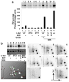
Fig. 2. MEK-1 expression induces increased C-Raf phosphorylation
(a) COS-7 cells were transfected with pMT2-myc-C-Raf wildtype (wt), S471A or S471T mutants alone (lanes 1–7) or together with pExchange 5a-FLAG-MEK-1 (lanes 8–11). Following 24 h, cells were deprived of serum for 18 h, metabolically labeled with 32P and treated with vehicle or 100 ng/ml EGF for 30 min. Indicated samples (lanes 7 and 8) were treated with 20 μM of the MEK inhibitor, U0126, 30 min prior to 32P-labeling. myc-C-Raf proteins were immunoprecipitated with a myc-epitope tag antibody and separated using SDS-PAGE. 32P incorporation in myc-C-Raf was visualized using a phosphor imager (insert) and quantified using the Bio-Rad Quantity One software. (b) Cells were transfected as indicated and the samples were analyzed as in a (left top panel). The 32P bands were excised from the membrane and 2D phosphopeptide mapping was performed as described in Materials and Methods (panels 1–8). 10% of the sample was used to examine myc-C-Raf recovery (bottom part). A schematic representation of the phosphopeptide spots, including the migration positions of known C-Raf phosphopeptides are depicted in panel 9. Arrows indicate the orientation of electrophoresis and TLC chromatography. Note that equal amounts of counts were loaded from each sample (1200 cpm, excluding sample 7) to allow comparison of phosphorylation stoichiometry between the samples. For sample 7, S259/621A mutant, only 300 cpm were available since this mutant is consistently less phosphorylated than the other C-Raf forms. The results are representative of three independent experiments.
Fig. 3
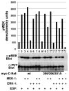
Fig. 3. MEK-1 expression increases C-Raf kinase activity
COS-7 cells were transfected with pMT2-myc-C-Raf (wt) or S289/296/301A mutant alone or together with pExchange 5a-FLAG-MEK-1 or pMT2-HA-ERK1 as indicated. After 24 h, cells were deprived of serum for 18 h and treated with vehicle or 100 ng/ml EGF for 20 min. C-Raf kinase activity in myc-immunoprecipitates was assayed using recombinant GST-MEK-1 as a substrate as described in Materials and Methods. Presented are phospho-MEK and myc immunoblots showing MEK phosphorylation and myc-C-Raf recovery, respectively. The bar graph shows densitometry quantification of the phospho-GST-MEK-1 band. Presented are representative results of three independent experiments.
Fig. 4
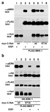
Fig. 4. Coexpression of MEK-1 and C-Raf induces constitutive activation of MEK and ERK
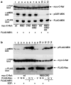
(a) COS-7 cells were transfected with wildtype pMT2-myc-C-Raf (wt, lanes 1,2 and 5,6), the S471A mutant (471A, lanes 7,8) or empty pMT2 vector (lanes 3,4), alone (lanes 1,2) or together with pExchange 5a-FLAG-MEK-1 (lanes 3–8). After 24 h, cells were deprived of serum for 18 h and treated with vehicle or 100 ng/ml EGF for 20 min as indicated. MEK-1 activation in cells was determined following FLAG immunoprecipitation and immunoblotting with phospho-MEK (top panel). MEK-1 recovery was examined by FLAG-immunoblotting (middle panel) and myc-C-Raf expression was determined in cell extracts by myc immunoblotting (bottom panel). (b) COS-7 cells expressing the above C-Raf and MEK-1 expression vectors, as indicated, together with pMT2-HA-ERK-1 vector were stimulated with EGF as in a. ERK phosphorylation was determined following HA immunoprecipitation (top panel). ERK recovery was determined using HA immunoblotting. MEK and Raf expressions were determined in cell lysates using FLAG and myc immunoblotting respectively. Presented are representative results of five independent experiments.
Fig. 5
Fig. 5. Small increases in MEK-1 and C-Raf expression are sufficient for ERK activation
(a) COS-7 cells were transfected with the indicated amounts of pExchange 5a-FLAG-MEK-1 and pMT2-HA-ERK-1 together with increasing amounts of pMT2-myc-C-Raf (0.01 – 1 μg/plate). After 24 h, cells were deprived of serum for 18 h and treated with vehicle or 100 ng/ml EGF for 20 min, where indicated. ERK-1 activation in cells was determined by immunoblotting for phospho-ERK (top panel). Expression of myc-C-Raf, FLAG-MEK and HA-ERK was examined by myc, FLAG and HA immunoblotting, respectively. Note that increased expression of myc-C-Raf resulted in decreased HA-ERK expression. This was probably due to DNA interference since both use the pMT2 backbone. Empty pMT2 vector was used to adjust for total DNA amounts in these transfections. (b and c) COS-7 cells were transfected with pMT2-myc-C-Raf and the indicated amounts of pMT2-HA-ERK-1 together with increasing amounts of pExchange 5a-FLAG-MEK-1 (0.01–1 μg/plate). After 24 h, cells were deprived of serum for 18 h and C-Raf kinase activity was examined in vitro in myc immunoprecipitates using recombinant GST-MEK-1 as a substrate and 32P-ATP (b, top panel). myc-C-Raf recovery, MEK and ERK expressions are also provided (b). Quantification of GST-MEK phosphorylation is provided in c. Presented are representative results of three independent experiments.
Fig. 6
Fig. 6. MEK-induced C-Raf activation is Ras-independent
(a) COS-7 cells were transfected with pExchange 5a-FLAG-MEK-1 alone (lanes 1,2) or together with pMT2-myc-C-Raf (wt) or K84A/L86A/K87A (RBD, Ras binding domain), C165/168S (CRD, cysteine-rich domain) C-Raf mutants or with same C-Raf forms containing a c-terminal CaaX motif for membrane targeting (caax). After 24 h, cells were deprived of serum for 18 h and treated with vehicle or with 100 ng/ml EGF for 20 min as indicated. Presented are a pMEK immunoblot of FLAG-immunoprecipitates, a MEK immunoblot showing MEK recovery and a myc immunoblot showing the expression of the myc-C-Raf variants. (b) COS-7 cells were transfected as in a with the indicated DNA combinations of MEK, C-Raf variants and pMT2-HA-ERK. Presented are a pERK immunoblot in cell extracts and a myc immunoblot showing the expression of the myc-C-Raf variants. CRD/SA is C165/168A mutant, RBD/KD and RBD/CRD/KD are RBD and RBD/CRD mutants, respectively, that include also the K375M mutation in the ATP binding pocket that abrogates C-Raf kinase activity (see materials and methods for more details). Presented are representative results of four independent experiments.
Fig. 7
Fig. 7. Additive C-Raf activation by MEK and Ras
(a) COS-7 cells were transfected with pMT2-myc-C-Raf (wt) or K84A/L86A/K87A (RBD), C165/168S (CRD), K375M (KD) or their indicated combinations, alone or with pExchange 5a-FLAG-MEK-1 as indicated. After 24 h, cells were deprived of serum for 18 h and C-Raf kinase activity was determined in myc-immunoprecipitates using an in vitro kinase assay with recombinant GST-MEK as a substrate. Presented are a myc immunoblot showing myc-C-Raf recovery (top panel) pMEK immunoblot of the GST-MEK substrate (middle panel) and FLAG immunoblot of cell extracts showing FLAG-MEK expression in the cells (bottom panel). (b) COS-7 cells were transfected with pMT2-myc-C-Raf (wt) or the RBD/CRD mutant together with pExchange 5a FLAG-MEK-1 and pCMV5-FLAG-Ras V12 (V, constitutively active) or N17 (N, inactive mutant) as indicated. After 24 h, cells were deprived of serum for 18 h and treated with vehicle or with 100 ng/ml EGF for 20 min. Presented are a pMEK immunoblot of cell extracts (top panel), a myc immunoblot showing the expression of myc-C-Raf variants (middle panel) and a FLAG immunoblot showing the expression of the FLAG-Ras variants (bottom panel). The results are representative of two independent experiments.
Fig. 8
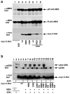
Fig. 8. MEK kinase activity is not required for the MEK-induced C-Raf activation
(a) COS-7 cells were transfected with pMT2-myc-C-Raf together with pMT2-HA-ERK-1 and pExchange 5a-FLAG-MEK-1 wildtype (wt) or a kinase dead mutant (KD, K97M) as indicated. After 24 h, cells were deprived of serum for 18 h and treated with vehicle or 100 ng/ml EGF for 20 min. Presented are phospho-MEK and phospho-ERK immunoblots of cell lysates showing MEK and ERK phosphorylation, respectively and FLAG, HA and myc-immunoblots showing FLAG-MEK-1, HA-ERK-1 and myc-C-Raf expression. (b) COS-7 cells were transfected with pMT2-myc-C-Raf together with pExchange 5a-FLAG-MEK-1 (wt, lanes 7,8) or K97M mutant (KD, lane 1) as indicated. After 24 h, cells were deprived of serum for 18 h in the presence or absence of the heat-shock protein 90 inhibitor 17-AAG (1 μM) and treated with vehicle or 100 ng/ml EGF for 20 min. C-Raf kinase activity was measured in vitro in myc-immunoprecipitates using a coupled kinase assay with recombinant ERK-1 serving as a substrate. Presented are phospho-ERK and myc immunoblots of the kinase reaction showing Raf kinase activity and myc-C-Raf recovery and phospho-MEK and FLAG immunoblots of cell extracts showing MEK phosphorylation and expression in cells. Alpha-tubulin immunoblot shows equal protein loading. Note that 17-AAG treatment results in reduced myc-C-Raf and FLAG-MEK protein expressions (lanes 4,6,8) as has been previously reported. (c) COS-7 cells were transfected with pMT2-myc-C-Raf (wt) or S338/339A mutant together with pMT2-HA-ERK-1 and pExchange 5a-FLAG-MEK-1 wildtype (wt) or a proline-rich domain deletion mutant (Δ, deletion of amino acids 265-301) as indicated. After 24 h, cells were deprived of serum for 18 h and treated with vehicle or 100 ng/ml EGF for 20 min. Following protein extraction, samples were split and half was used to immunoprecipitate MEK using FLAG antibody (left part) and half to immunoprecipitate Raf using myc antibody (right part). In the left part, presented are FLAG, myc and pS296 C-Raf immunoblots in FLAG-immunoprecipitates (FLAG-IP), showing FLAG-MEK, myc-C-Raf and phospho-S296 C-Raf recoveries, respectively, and a pERK immunoblot of cell extracts, showing ERK phosphorylation in cells. In the right part, presented are pS338 C-Raf and myc immunoblots in myc-immunoprecipitates (myc-IP), showing C-Raf phosphorylation at the S338 site and myc-C-Raf recovery. Note that the Δ 265-301 MEK-1 mutant activates and binds C-Raf as efficiently as full-length MEK-1.
Fig. 9
Fig. 9. A model for Ras-independent C-Raf-MEK activation
In normal conditions C-Raf and MEK expressions are low and they are maintained apart by cellular factors such as RKIP and MST2 (a). In certain conditions, such as cancer, increased expression of Raf and MEK (b) or down regulation of the sequestering factors (c) allows increased C-Raf-MEK binding, resulting in C-Raf hyper-phosphorylation and activation.

References

    1. Leicht DT, Balan V, Kaplun A, Singh-Gupta V, Kaplun L, Dobson M, Tzivion G. Raf kinases: function, regulation and role in human cancer. Biochim Biophys Acta. 2007;1773:1196–1212. - PMC - PubMed
    1. Roskoski R., Jr RAF protein-serine/threonine kinases: structure and regulation. Biochem Biophys Res Commun. 2010;399:313–317. - PubMed
    1. Udell CM, Rajakulendran T, Sicheri F, Therrien M. Mechanistic principles of RAF kinase signaling. Cell Mol Life Sci. 2011;68:553–565. - PMC - PubMed
    1. Matallanas D, Birtwistle M, Romano D, Zebisch A, Rauch J, von Kriegsheim A, Kolch W. Raf family kinases: old dogs have learned new tricks. Genes Cancer. 2011;2:232–260. - PMC - PubMed
    1. Avruch J, Zhang XF, Kyriakis JM. Raf meets Ras: completing the framework of a signal transduction pathway. Trends Biochem Sci. 1994;19:279–283. - PubMed

Publication types

MeSH terms

Substances

LinkOut - more resources