. 2013:4:1399.
doi: 10.1038/ncomms2406.

In vitro fabrication of functional three-dimensional tissues with perfusable blood vessels

Affiliations
Free PMC article

In vitro fabrication of functional three-dimensional tissues with perfusable blood vessels

Hidekazu Sekine et al. Nat Commun. 2013.
Free PMC article

Abstract

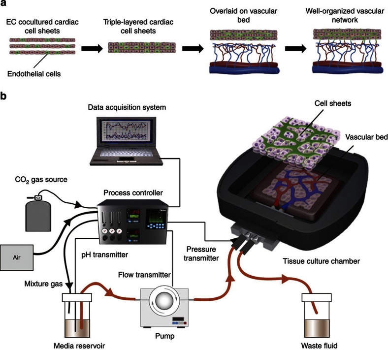
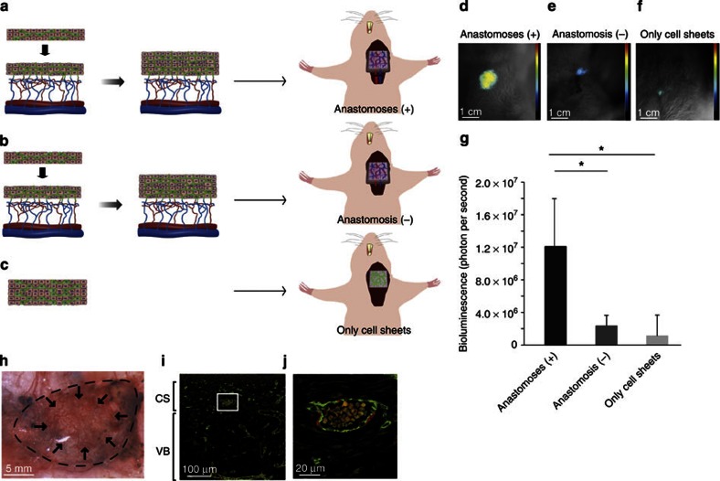
In vitro fabrication of functional vascularized three-dimensional tissues has been a long-standing objective in the field of tissue engineering. Here we report a technique to engineer cardiac tissues with perfusable blood vessels in vitro. Using resected tissue with a connectable artery and vein as a vascular bed, we overlay triple-layer cardiac cell sheets produced from coculture with endothelial cells, and support the tissue construct with media perfused in a bioreactor. We show that endothelial cells connect to capillaries in the vascular bed and form tubular lumens, creating in vitro perfusable blood vessels in the cardiac cell sheets. Thicker engineered tissues can be produced in vitro by overlaying additional triple-layer cell sheets. The vascularized cardiac tissues beat and can be transplanted with blood vessel anastomoses. This technique may create new opportunities for in vitro tissue engineering and has potential therapeutic applications.


Figures

Figure 1
Figure 1. In vitro engineering of functional 3-D tissue with perfusable blood vessels.
(a) To engineer cell sheet constructs with perfusable blood vessels, EC cocultured cardiac cell sheets are stacked, and then overlaid on a vascular bed in vitro. After appropriate perfusion using a bioreactor, the cocultured ECs formed new blood vessels and connected with the blood vessels that originated from the vascular bed. Finally, the cell sheet constructs survive via the media supplied through the new vessels that formed in vitro. (b) The engineered constructs are perfused in a custom-made bioreactor. The bioreactor is a one-pass system consisting of a delivery pump, a custom-made tissue culture chamber, pH transmitter, flow transmitter, pressure transmitter, CO2 gas source, process controller and data acquisition system. The construct is placed in the tissue culture chamber to become a vascular bed and the femoral vessels are connected to two micro polyurethane tubes for culture media perfusion. The flow is generated by a delivery pump from the media reservoir monitored by a pressure sensor positioned on the arterial inlet side, passing through the tissue culture chamber to exit into a waste fluid tank. The pH is measured and controlled via a process controller to verifying the concentration of CO2 gas. The pH, temperature and arterial pressure sensors are connected to the computer via a process controller.
Figure 2
Figure 2. Perfusable blood vessel formation and viable cardiac tissue fabrication.
Blood vessel formation and tissue viability were evaluated among the four groups, with and without EC coculture, and with or without FGF-2 administration in the perfusion medium. Each group was indicated as EC (−) FGF (−), EC (−) FGF (+), EC (+) FGF (−) and EC (+) FGF (+). (ad) HE staining after a 3-day perfusion culture. Neither a luminal structure nor perfused black ink were detected in either the EC (−) FGF (−) group or in the EC (−) FGF (+) group. In the EC (+) FGF (−) group, a luminal structure was observed, but the black ink was hardly detected within it. In the EC (+) FGF (+) group, the infused black ink thoroughly reached the blood vessel structure within cell sheet layers (red arrows). (eh) CD31 staining for ECs. No blood vessel structure was observed within the cell sheet layers in either the EC (−) FGF (−) group or the EC (−) FGF (+) group. Significant tubular blood vessel structures (white arrows) were observed in the EC (+) FGF (−) group and in the EC (+) FGF (+) group. cell sheet (CS) indicates cell sheet constructs and vascular bed (VB) indicates vascular beds. (io) Luciferase-expressing triple-layer cell sheets were overlaid on the vascular beds. After continuous luciferin infusion, serial bioluminescent imaging of each group was analysed. The images captured at 45 min are shown (il). Representative dynamic tracing is demonstrated (m). The bioluminescent intensity at 45 and 90 min is shown (n,o). The intensity increased in the early phase in the EC (+) FGF (−) group and the EC (+) FGF (+) group. On the other hand, it increased in the late phase in the EC (−) FGF (−) group and the EC (−) FGF (+) group. FGF-2 accelerated the increasing rate, regardless of EC coculture. Data are shown as mean±s.d. (*P<0.05) (n=4). (p) Sarcomeric α-actinin staining (red) with nuclei staining (blue) revealed well-differentiated sarcomeres within the cell sheet construct in the EC (+) FGF (+) group. (q) HE staining showed that perfused black ink is occasionally detected EC(+) FGF(−) group.
Figure 3
Figure 3. Contribution of cocultured ECs within the construct.
(a) Scanning two-dimensional fluorescent images of the constructs containing cocultured green fluorescence protein (GFP)-expressing ECs were obtained after the constructs were perfused with red fluorescent dextran. Three xy images at different depths of the cell sheet (CS) regions (b, e and h), border region (c, f and i) and vascular bed (VB) region (d, g and j) are presented. (bd) Without FGF-2 treatment, the GFP-positive EC networks remain within the cell sheets, but dextran is hardly observed within the cell sheets. (eg) In the case of FGF-2-treated constructs, red fluorescent dextran passed through the GFP-positive EC tubular structures. (hj) When 4-μm-diameter red fluorescent spheres were perfused, many microspheres were observed in the GFP-positive blood vessels of the cell sheets when treated with FGF-2. (k) Immunostaining with CD31 (red: all ECs) and GFP (green: ECs originated from the cell sheet) demonstrated that cocultured ECs migrated into the vascular bed and connected to the blood vessels within the bed. An arrow indicates the communication between migrating ECs originated from the cell sheet and vascular bed originated ECs. (l) Fused blood vessels containing both vascular bed (red) and cell sheet (green)-derived ECs were present in the vascular bed. (m) By contrast, when GFP-negative cell sheets were overlaid on the GFP-expressing vascular bed, GFP-positive cells never migrated into the cell sheet layers.
Figure 4
Figure 4. Double-step overlaying of triple-layer cell sheets and viability assay.
(a) The double-step overlaying is schematically illustrated. Triple-layer cocultured cardiac cell sheets are overlaid on the vascular bed and perfused. After 3 days of cultivation, to provide sufficient vascularization within the first graft, the second triple-layer tissue is overlaid on the first graft and the construct is perfused for a further 3 days. Finally, the whole construct is thoroughly vascularized. (b) The single-step overlaying with six-layer cell sheets is schematically illustrated. Six-layer cell sheet tissues are overlaid, in one step, on the vascular bed and perfused for 6 days. (c,d) HE and Azan staining for blood-perfused double-step constructs showed that blood cells well reached the six-layer cell sheets (white arrows) and that thicker and more cell-dense tissue was successfully fabricated. (e,f) HE and Azan staining for blood cell-perfused one-step constructs shows some necrotic cells (black arrow heads) and relatively cell-sparse tissues. (gi) After continuous luciferin infusion, serial bioluminescent imaging of each group was analysed. The images captured at 60 min are shown (g,h). The intensity from the double-step procedure was significantly higher than that from the single-step procedure (i). Data are shown as mean±s.d. (*P<0.05) (n=4). Triple-layer cell sheets are overlaid four times on the vascular bed to produce a thick 12-layer construct (j).
Figure 5
Figure 5. Transplantation of in vitro vascularized cardiac tissues.
(a) The six-layer tissue graft engineered by the double-step procedure was removed from the bioreactor system and transplanted in the neck of a nude rat with anastomosis between the protruding femoral vessels of the graft and the connectable neck blood vessels of the host rat. (b) The six-layer graft was transplanted without the vessel anastomosis. (c) The six-layer cell sheets (CS) without the vascular bed (VB) were simply transplanted. (df) In vivo bioluminescent imaging of each graft is shown. Obvious signals were observed in the graft with vessel anastomoses (d). The intensity was low in the graft without anastomosis (e) and in the graft without a vascular bed (f). (g) The intensity was significantly the highest in the graft with vessel anastomosis, compared with the other two grafts. Data are shown as mean±s.d. (*P<0.05) (n=4). (h) The vascularized graft with blood vessel anastomoses also maintained its vascular structure at 2 weeks after the procedure. Dotted line indicates the area of the graft with the vascular bed and arrows indicate the area of the cell sheet layers. (i) Immunostaining for CD31 (green: ECs) and calponin (red: smooth muscle cell) shows the uniform distribution of micro blood vessels in the cell sheet construct. (j) The detail in the white box of i illustrates the characteristics of a matured blood vessels including red blood cells.

References

    1. Lango A.H. et al. Hundreds of variants clustered in genomic loci and biological pathways affect human height. Nature 467, 832–838 (2010) . - PMC - PubMed
    1. Langer R. & Vacanti J. P.. Tissue engineering. Science 260, 920–926 (1993) . - PubMed
    1. Cao Y., Vacanti J. P., Paige K. T., Upton J. & Vacanti C. A.. Transplantation of chondrocytes utilizing a polymer-cell construct to produce tissue-engineered cartilage in the shape of a human ear. Plast. Reconstr. Surg. 100, 297–302 (1997) . - PubMed
    1. Kirsner R. S., Falanga V. & Eaglstein W. H.. The development of bioengineered skin. Trends Biotechnol. 16, 246–249 (1998) . - PubMed
    1. Brittberg M. et al. Treatment of deep cartilage defects in the knee with autologous chondrocyte transplantation. N. Engl. J. Med. 331, 889–895 (1994) . - PubMed

Publication types