. 2013:4:1406.
doi: 10.1038/ncomms2420.

A point mutation in Semaphorin 4A associates with defective endosomal sorting and causes retinal degeneration

Affiliations
Free PMC article

A point mutation in Semaphorin 4A associates with defective endosomal sorting and causes retinal degeneration

Satoshi Nojima et al. Nat Commun. 2013.
Free PMC article

Abstract

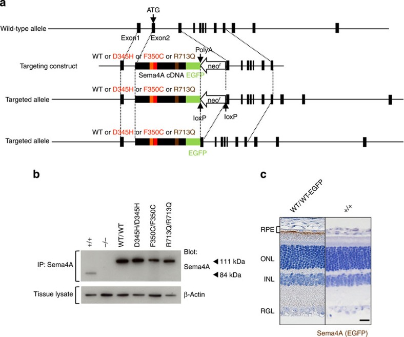
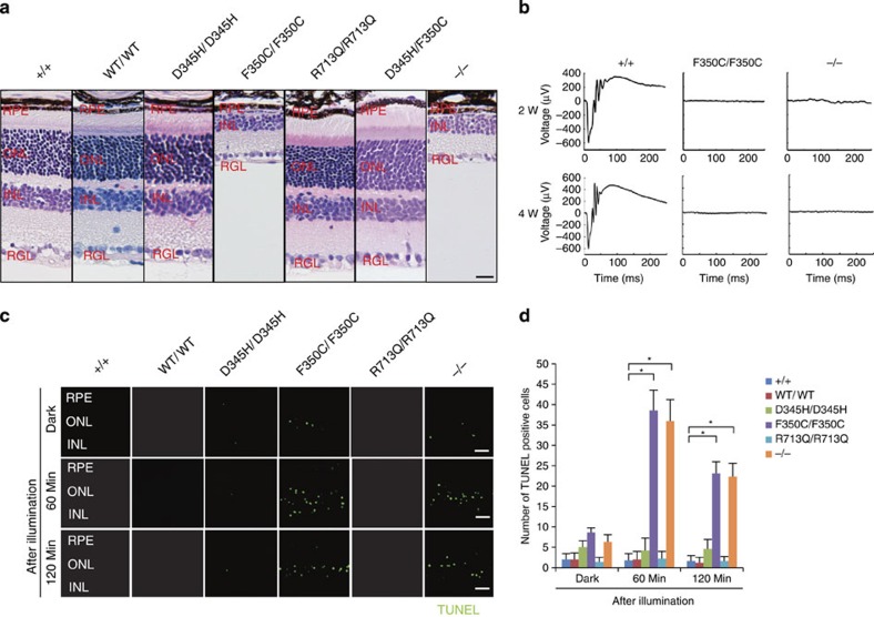
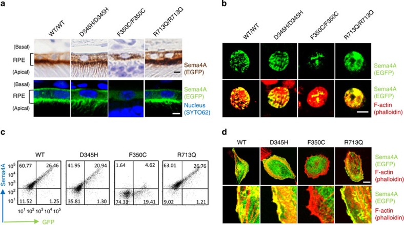
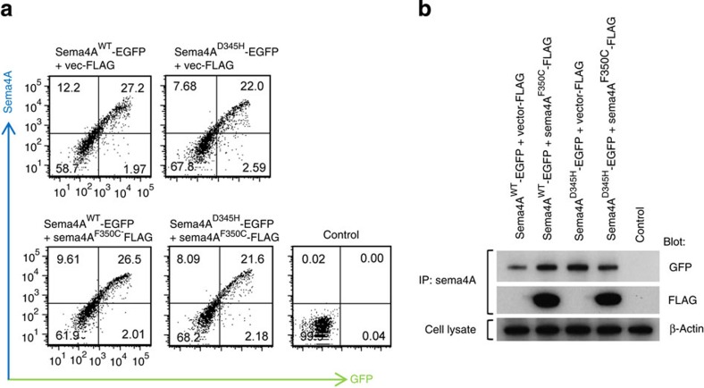
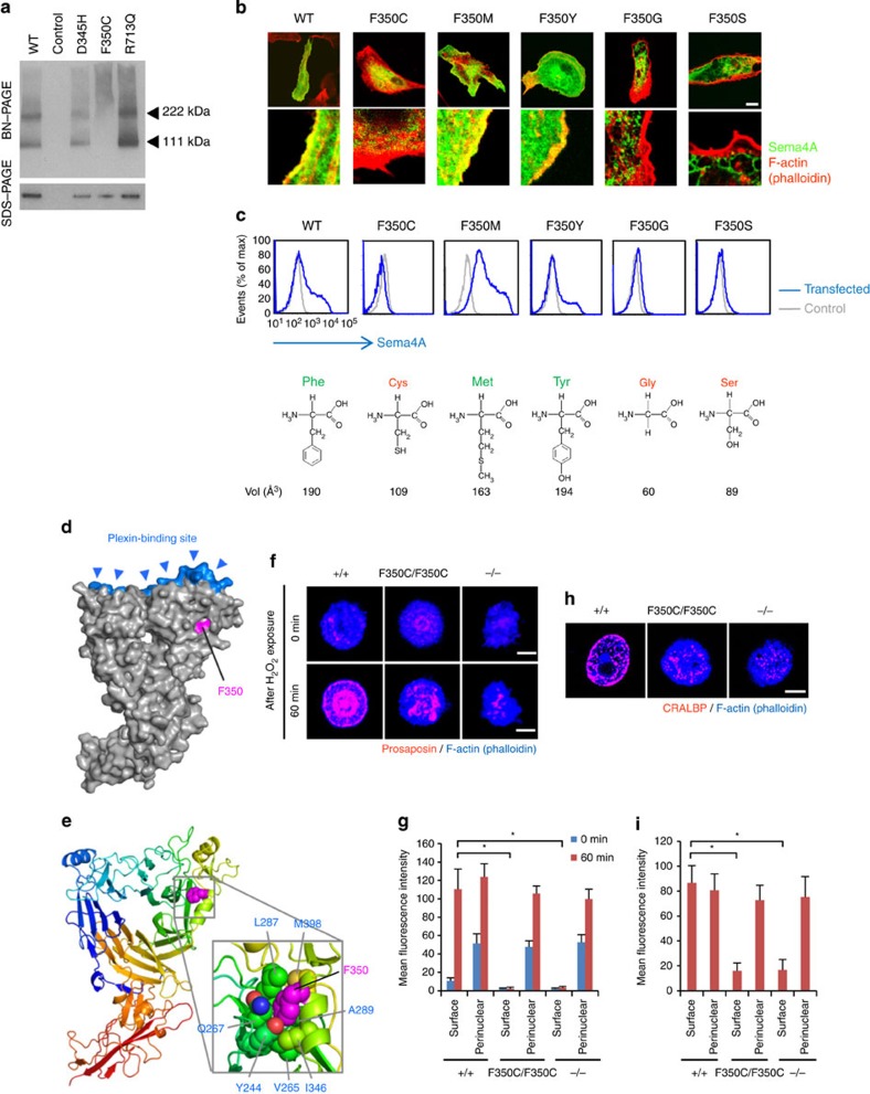
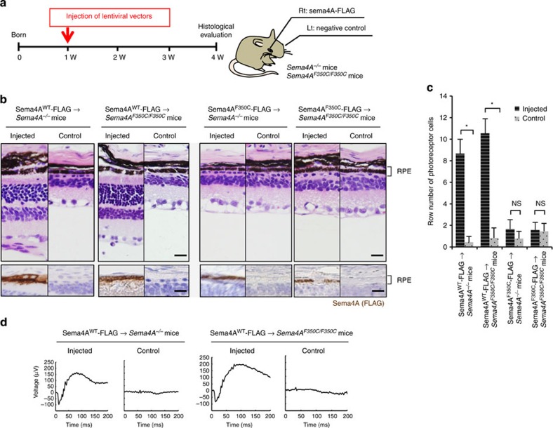
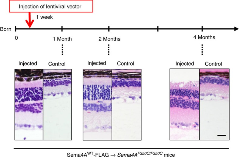
Semaphorin 4A (Sema4A) has an essential role in photoreceptor survival. In humans, mutations in Sema4A are thought to contribute to retinal degenerative diseases. Here we generate a series of knock-in mouse lines with corresponding mutations (D345H, F350C or R713Q) in the Sema4A gene and find that Sema4A(F350C) causes retinal degeneration phenotypes. The F350C mutation results in abnormal localization of the Sema4A protein, leading to impaired endosomal sorting of molecules indispensable for photoreceptor survival. Additionally, protein structural modelling reveals that the side chain of the 350th amino acid is critical to retain the proper protein conformation. Furthermore, Sema4A gene transfer successfully prevents photoreceptor degeneration in Sema4A(F350C/F350C) and Sema4A(-/-) mice. Thus, our findings not only indicate the importance of the Sema4A protein conformation in human and mouse retina homeostasis but also identify a novel therapeutic target for retinal degenerative diseases.


Figures

Figure 1
Figure 1. Generation of knock-in mice.
(a) Schematic diagram of the endogenous mouse locus for the Sema4A knock-in vectors and the resulting Sema4A proteins after homologous recombination. Full-length cDNA fragments of WT Sema4A or mutated Sema4A (D345H, F350C or R713Q) fused with EGFP at the C-terminus were inserted into exon 2 and exon 3 of the Sema4A gene. The neomycin resistance gene was flanked by loxP sites so that it could be excised upon expression of Cre recombinase. The gene structure of the WT Sema4A allele (top), Sema4A-targeting construct (second row) and the resulting Sema4A-targeted allele in which the neomycin resistance gene was (bottom) or was not (third row) excised. (b) Expression of Sema4A proteins in the brain tissues of knock-in mice. Brain tissues from WT (Sema4A+/+), Sema4AWT/WT, Sema4AD345H/D345H, Sema4AF350C/F350C, Sema4AR713Q/R713Q and Sema4A−/− (negative control) mice were lysed and subjected to immunoprecipitation (IP) and Western blot analyses using an anti-Sema4A antibody. The 111-kDa bands represent the mutant Sema4A-EGFP proteins (EGFP-tagged), while the 84-kDa band represents the endogenous wild-type Sema4A protein. All series of knock-in mice expressed sufficient amounts of Sema4A protein. (c) Paraffin sections of Sema4AWT/WT or wild-type (Sema4A+/+) (negative control) retinas were examined by immunohistochemistry with an anti-GFP antibody. Sema4A normally localizes at the apical surface of RPE cells in the retina. Scale bar, 50 μm.
Figure 2
Figure 2. Light-induced photoreceptor damage in retinas of Sema4AF350C/F350C mice.
(a) Hematoxylin and eosin (HE) staining of retinas in 4-week-old wild-type (Sema4A+/+), Sema4AWT/WT, Sema4AD345H/D345H, Sema4AF350C/F350C, Sema4AR713Q/R713Q, Sema4AD345H/F350C and Sema4A-deficient (Sema4A−/−) mice. Among them, Sema4AF350C/F350C and Sema4A−/− retinas showed loss of the outer nuclear layer. Scale bar, 50 μm. RPE, retinal pigment epithelium; ONL, outer nuclear layer; INL, inner nuclear layer; RGL, retinal ganglion layer. (b) ERG responses to single flashes were recorded using wild-type (Sema4A+/+), Sema4AF350C/F350C and Sema4A−/− mice in a scotopic condition at 2 or 4 weeks of age. Virtually no ERG responses were detected in Sema4A−/− and Sema4AF350C/F350C retinas as early as 2 weeks of age. (c) Representative images from the TUNEL assay using P10 mouse retinas after 0, 60 and 120 min of light exposure. Scale bar, 50 μm. (d) Histogram showing the average number of TUNEL-positive cells (±s.e.m.; n=5–10) in retinas. *P<0.01 (Student’s t-test). Photoreceptor apoptosis peaked in Sema4A−/− and Sema4AF350C/F350C retinas after 60 min of exposure. Data are representative of three independent experiments.
Figure 3
Figure 3. The Sema4AF350C proteins are mis-localized in RPE cells.
(a) Representative images of immunostaining of EGFP-tagged Sema4A proteins in RPE cells in Sema4AWT/WT, Sema4AD345H/D345H, Sema4AF350C/F350C and Sema4AR713Q/R713Q retinas using DAB (top) or fluorescence (bottom). EGFP is shown in brown (top) or green (bottom), and nuclei were visualized with SYTO 62 staining as shown in blue (bottom). Fluorescent signal of EGFP was enhanced by immunostaining using anti-GFP and Alexa Fluor 488-conjugated secondary antibodies (bottom). Scale bar, 2 μm. (b) Immunofluorescent images of primary cultured RPE cells derived from Sema4AWT/WT, Sema4AD345H/D345H, Sema4AF350C/F350C and Sema4AR713Q/R713Q mice. EGFP was stained with anti-GFP and Alexa Fluor 488-conjugated secondary antibodies to enhance the GFP signals (green), and the cytoskeleton was visualized by staining with Alexa Fluor 546-conjugated phalloidin as shown in red. Scale bar, 5 μm. (c) Expression of Sema4A on the plasma membrane. COS-7 cells were transfected with plasmid constructs encoding Sema4AWT-EGFP, Sema4AD345H-EGFP, Sema4AF350C-EGFP and Sema4AR713Q-EGFP and incubated for 48 h. Subsequently, the cells were stained with an anti-Sema4A antibody and analysed by flow cytometry. Data are representatives of three experiments. (d) ARPE-19 cells were transfected with plasmid constructs expressing Sema4AWT-EGFP, Sema4AD345H-EGFP, Sema4AF350C-EGFP and Sema4AR713Q-EGFP, incubated for 48 h, fixed, stained with Alexa Fluor 546-conjugated phalloidin, and then examined by confocal microscopy. Representative (top) and enlarged images (bottom) are shown. Scale bar, 10 μm.
Figure 4
Figure 4. Sema4AF350C protein does not function in a dominant-negative manner.
(a) COS-7 cells were transfected with plasmid constructs encoding Sema4AWT-EGFP, Sema4AD345H-EGFP with or without co-transfection of Sema4AF350C-FLAG. After incubation for 48 h, the cells were stained with an anti-Sema4A antibody and analysed by flow cytometry. Control represents untransfected COS-7 cells with staining in the same conditions. (b) The same transfected COS-7 cells were lysed and subjected to immunoprecipitation (IP) with an anti-Sema4A antibody and subsequent Western blot analyses using anti-GFP (represents Sema4AWT-EGFP or Sema4AD345H-EGFP) or anti-FLAG (represents Sema4AF350C-FLAG) antibodies. Every Sema4A protein was sufficiently expressed in COS-7 cells.
Figure 5
Figure 5. The Sema4AF350C proteins exhibit severe structural defects and impaired function.
(a) BN-PAGE and SDS–PAGE with an anti-Sema4A antibody were performed using cell lysates derived from COS-7 cells transfected with constructs expressing Sema4A-EGFP mutant proteins, or pEGFP vector (negative control). In BN-PAGE, the 222-kDa bands represent Sema4A dimers, while the 111-kDa bands represent Sema4A monomers. SDS–PAGE was performed after immunoprecipitation with an anti-Sema4A antibody, using the same lysate as BN-PAGE. (b) Representative images (top) and enlarged images (bottom) obtained by confocal microscopy. ARPE-19 cells were transfected with the plasmid constructs expressing Sema4AF350-EGFP mutant proteins, incubated for 48 h and stained with phalloidin. Scale bar, 10 μm. (c) COS-7 cells were transfected with plasmid constructs and incubated for 48 h, stained with an anti-Sema4A antibody and analysed by flow cytometry. Structural diagrams for amino-acid residue replaced with F350 in each mutant are shown below the histograms, together with their apparent volume per molecule in Å3 (ref. 34). (d,e) Structural modelling of mouse Sema4A ectodomain, which was built using the structure of human Sema4D structural model previously reported. (f) Immunofluorescent images of mouse RPE cells after H2O2 treatment (250 μM). Prosaposin (red) was peripherally distributed in wild-type (Sema4A+/+) RPE cells but not Sema4A−/− or Sema4AF350C/F350C RPE cells. Scale bar, 5 μm. (g) Quantitative analysis of the normalized fluorescence intensity of prosaposin at the surface or perinuclear area of the respective RPE cells with (after 60 min) or without (0 min) H2O2 treatment (±s.e.m.; n=10). To quantify the intensity, we calculated the mean normalized intensity within the square with its side having an outer 1/6 of radius (‘surface’) or inner 1/6–3/6 of radius (‘perinuclear’). *P<0.01 (Student’s t-test). (h) Immunofluorescent images of mouse RPE cells using an anti-CRALBP antibody. Scale bar, 5 μm. (i) Quantitative analysis of the normalized fluorescence intensity of CRALBP on the surface or perinuclear area of RPE cells (±s.e.m.; n=10–15). *P<0.01 (Student’s t-test).
Figure 6
Figure 6. Sema4A gene transfer prevents photoreceptor degeneration in the retinas of Sema4A−/− and Sema4AF350C/F350C mice.
(a) Schematic diagram of the protocol: At 1 week of age, suspensions of lentiviral vectors expressing Sema4AWT-FLAG or Sema4AF350C-FLAG were injected into the subretinal space of Sema4A−/− or Sema4AF350C/F350C infant mice. Viral suspensions were injected into the right eye, while the left eye was used as a negative control (only eyelids were incised). At 4 weeks of age, their eye tissues were fixed and sectioned. (b) (Top) Haematoxylin and eosin (HE) staining of the retinal sections from each mouse. Scale bar, 50 μm. (Bottom) The serial sections from those of HE staining were examined by immunohistochemistry (IHC) using an anti-FLAG antibody. Scale bar, 50 μm. (c) Histogram showing the average number of photoreceptor cells (±s.e.m.; n=9–18) in retinas. *P<0.01 (Student’s t-test); NS, not significant. The number of row of photoreceptor cells was counted at three random points per retinal section in which Sema4AWT-FLAG or Sema4AF350C-FLAG was expressed in immunohistochemistry using an anti-FLAG antibody. (d) ERG responses to single flashes were recorded using Sema4A−/− or Sema4AF350C/F350C mice after Sema4A gene transfer. A suspension of lentiviral vectors expressing Sema4AWT-FLAG was injected into the retinas of Sema4A−/− or Sema4AF350C/F350C mice at 1 week of age, and ERGs were recorded at 4 weeks of age. Viral suspensions were injected into the right eye, while the left eye was used as a negative control (only eyelids were incised).
Figure 7
Figure 7. Long-term prevention of photoreceptor degeneration after Sema4A gene transfer.
Similar to Figure 6, gene transfer with the lentiviral vectors expressing Sema4AWT-FLAG was performed in 1-week-old Sema4AF350C/F350C mice. Subsequently, we evaluated the retinal histology of these mice at 1, 2 or 4 months of age to estimate the duration of the therapeutic effects. As shown in the representative images, photoreceptor cells were preserved at least 4 months after gene transfer. Scale bar, 50 μm.

Similar articles

Cited by

References

    1. Pacione L. R. et al.. Progress toward understanding the genetic and biochemical mechanisms of inherited photoreceptor degenerations. Annu. Rev. Neurosci. 26, 657–700 (2003) . - PubMed
    1. Wright A. F. et al.. Photoreceptor degeneration: genetic and mechanistic dissection of a complex trait. Nat. Rev. Genet. 11, 273–284 (2010) . - PubMed
    1. Kolodkin A. L., Matthes D. J. & Goodman C. S.. The semaphorin genes encode a family of transmembrane and secreted growth cone guidance molecules. Cell 75, 1389–1399 (1993) . - PubMed
    1. Serini G. et al.. Class 3 semaphorins control vascular morphogenesis by inhibiting integrin function. Nature 424, 391–397 (2003) . - PubMed
    1. Neufeld G. & Kessler O.. The semaphorins: versatile regulators of tumour progression and tumour angiogenesis. Nat. Rev. Cancer 8, 632–645 (2008) . - PubMed

Publication types

MeSH terms

LinkOut - more resources