. 2013:4:1407.
doi: 10.1038/ncomms2361.

Computational identification of a transiently open L1/S3 pocket for reactivation of mutant p53

Affiliations
Free PMC article

Computational identification of a transiently open L1/S3 pocket for reactivation of mutant p53

Christopher D Wassman et al. Nat Commun. 2013.
Free PMC article

Abstract

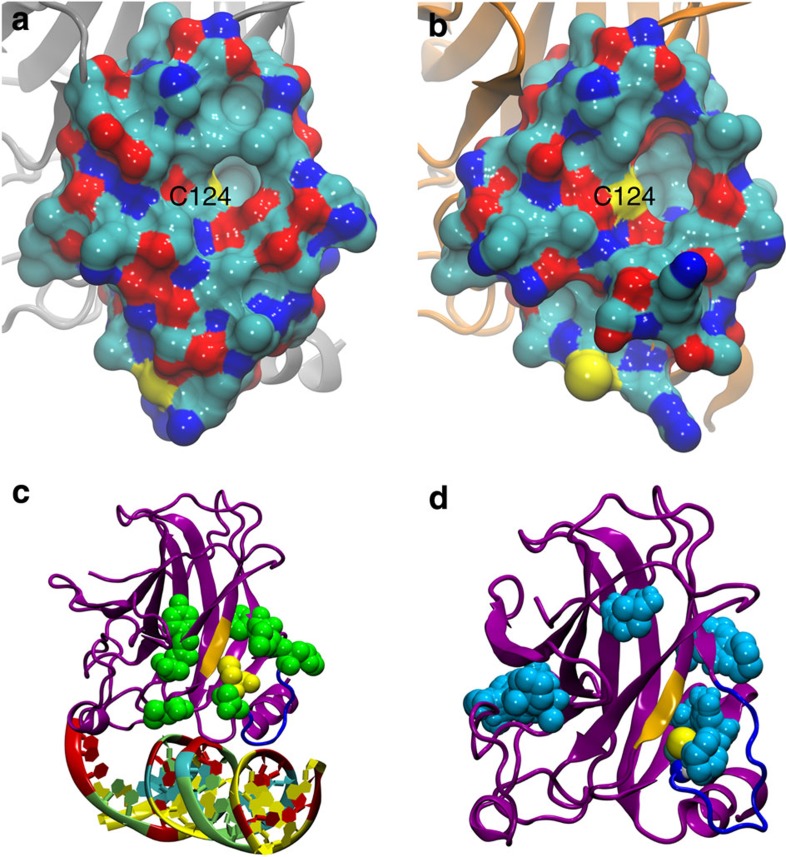
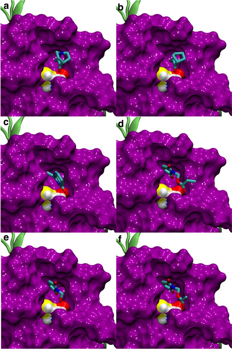
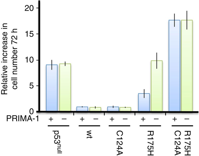
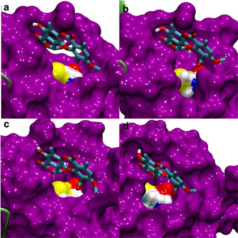
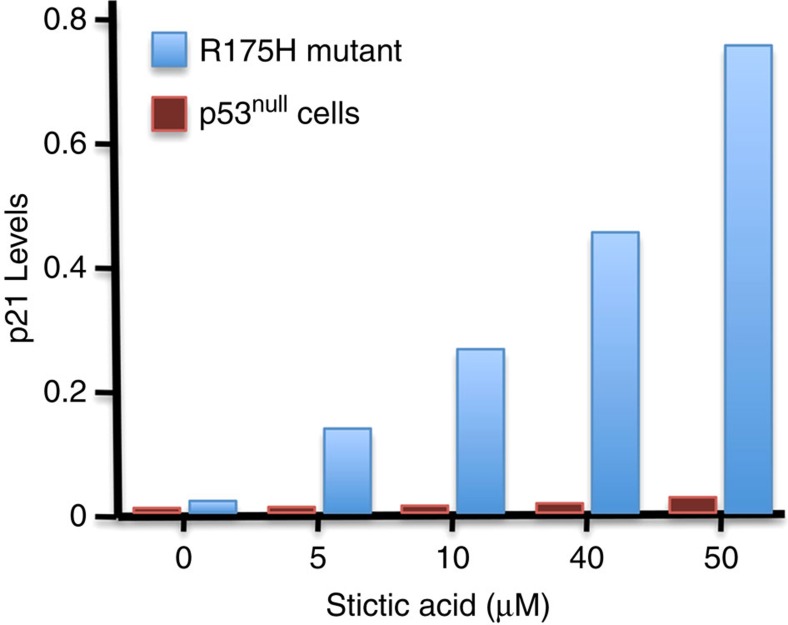
The tumour suppressor p53 is the most frequently mutated gene in human cancer. Reactivation of mutant p53 by small molecules is an exciting potential cancer therapy. Although several compounds restore wild-type function to mutant p53, their binding sites and mechanisms of action are elusive. Here computational methods identify a transiently open binding pocket between loop L1 and sheet S3 of the p53 core domain. Mutation of residue Cys124, located at the centre of the pocket, abolishes p53 reactivation of mutant R175H by PRIMA-1, a known reactivation compound. Ensemble-based virtual screening against this newly revealed pocket selects stictic acid as a potential p53 reactivation compound. In human osteosarcoma cells, stictic acid exhibits dose-dependent reactivation of p21 expression for mutant R175H more strongly than does PRIMA-1. These results indicate the L1/S3 pocket as a target for pharmaceutical reactivation of p53 mutants.


Figures

Figure 1
Figure 1. Molecular visualizations of the p53 core domain.
(a,b) All residues of the p53 core domain (1TSR-B33) within 10 Å of Cys124 are shown in a surface representation coloured by atom type: cyan, C; blue, N; red, O; yellow, S. (a) Cys124 is initially occluded in the wt crystal structure. (b) Structure extracted from MD simulations of mutant R273H reveals new breathing structural topography near Cys124 within the range of normal solution dynamics. (c,d) The p53 core domain is shown in magenta, loop L1 in blue, strand S3 in gold. (c) Cys124 (yellow) is surrounded by genetic mutation sites that can reactivate p53 function (green). (d) Druggable pockets (cyan) from FTMAP for the R273H mutant. The open L1/S3 pocket surrounds Cys124 (yellow).
Figure 2
Figure 2. Known p53 reactivation compounds docked into the open L1/S3 pocket of R175H.
(a) MQ (from PRIMA-1), (b) NB, (c) STIMA-1 (ref. 22), (d) MIRA-1 (ref. 19), (e) MIRA-2 (ref. 19), (f) MIRA-3 (ref. 19). Known p53 reaction compounds could dock favourably into the most-populated R175H MD trajectory cluster centroid that had an open L1/S3 pocket. All of the nine docking poses generated by Autodock Vina for each compound above scored within the published standard error of Autodock Vina docking scores (Supplementary Table S1). In each case, the binding pose depicted above had the smallest distance between the reactive methylene of the small molecule and the Cys124 sulphydryl group. The protein surface representation is purple, except Cys124 atoms are coloured by atom type as in Fig. 1a,b.
Figure 3
Figure 3. PRIMA-1 acts through cysteine 124 in p53.
p53null Saos-2 cells expressing wild-type p53, or the indicated p53 mutants, were treated with 50 μM preheated PRIMA-1 (blue bar) or the medium without PRIMA-1 (DMSO, green bar) for 72 h. The increase in cell number during 72 h of growth is shown. The s.d. represent four independent samples.
Figure 4
Figure 4. Stictic acid docked into open L1/S3 pocket of p53 variants.
Stictic acid could dock favourably into the open L1/S3 pocket of MD-generated snapshots for all four p53 variants studied: (a) wt p53; (b) R175H; (c) R273H; (d) G245S. Only polar hydrogens of stictic acid are depicted here. Colours are as in Fig. 2.
Figure 5
Figure 5. Dose-dependent p21 activation in response to stictic acid.
Saos-2 cells (p53null) and Saos-2 cells stably expressing the p53 cancer mutant R175H were treated with doxycycline for 8 h to induce expression of mutant R175H. Cells were then grown for 48 h in the presence of stictic acid at the concentrations indicated. p21 expression was quantified using a Fuji LAS-4000 imaging system and plotted normalized to the expression of tubulin in each sample.
Figure 6
Figure 6. p53-dependent activation of PUMA and p21 transcription reporter by reactivation compounds.
Preheated PRIMA-1 (25 μM), NSC-319425 (25 μM) and stictic acid (10 μM) were added to stable Soas-2 (p53null) cell lines harbouring (a) PUMA- or (b) p21-dependent luciferase reporter and expressing the p53 mutants R175H or G245S as indicated. Activation of PUMA and p21 transcriptional reporter was measured by luciferase activity assays. Reporter cell lines without p53 expression were processed in parallel and used for normalization to visualize p53-dependent effects. Data are represented as mean±s.d. (n=4).
Figure 7
Figure 7. Thermal stabilization of p53 mutants by reactivation compounds.
Selected concentrations of PRIMA-1 (blue, square), MQ (green, circle) and stictic acid (red, triangle) were added to 4 μM of wild type, G245S and R175H core domain protein. Thermal stabilization was measured using DSF. Data points are plotted as the mean of sextuplet measurements, with corresponding 95% confidence intervals. Confidence interval bars not visible are smaller than the symbol used to plot the data point. PRIMA-1 and MQ stabilized each p53 variant about 1 °C or less over this range of concentrations. Stictic acid resulted in the greatest stabilization for both p53 mutants studied.

References

    1. Olivier M. et al. The IARC TP53 database: new online mutation analysis and recommendations to users. Hum. Mutat. 19, 607–614 (2002) . - PubMed
    1. Soussi T. & Beroud C.. Assessing TP53 status in human tumours to evaluate clinical outcome. Nat. Rev. Cancer 1, 233–240 (2001) . - PubMed
    1. Joerger A. C. & Fersht A. R.. The tumor suppressor p53: from structures to drug discovery. Cold Spring Harb. Persp. Biol. 2, a000919 (2010) . - PMC - PubMed
    1. Martins C. P., Brown-Swigart L. & Evan G. I.. Modeling the therapeutic efficacy of p53 restoration in tumors. Cell 127, 1323–1334 (2006) . - PubMed
    1. Selivanova G. & Wiman K. G.. Reactivation of mutant p53: molecular mechanisms and therapeutic potential. Oncogene 26, 2243–2254 (2007) . - PubMed

Publication types

MeSH terms