

MUC1 marks collecting tubules, renal vesicles, comma- and S-shaped bodies in human developing kidney tubules, renal vesicles, comma- and s-shaped bodies in human kidney
- PMID: 23361236
- PMCID: PMC3567759
- DOI: 10.4081/ejh.2012.e40
MUC1 marks collecting tubules, renal vesicles, comma- and S-shaped bodies in human developing kidney tubules, renal vesicles, comma- and s-shaped bodies in human kidney
Abstract
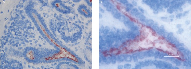
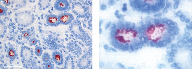
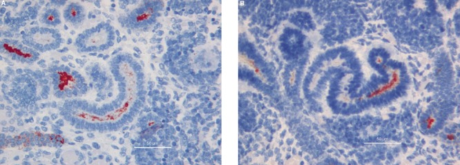
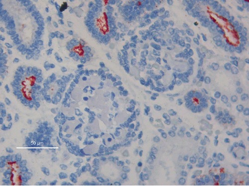
MUC1 is a transmembrane glycoprotein, apically expressed in most epithelial cells, used in the differential diagnosis of carcinomas and for discrimination of tumors of non-epithelial origin showing epithelioid features. Little attention has been paid so far though, on its possible significance in embryonic tissues. A preliminary study from our group revealed MUC1 expression in the cap mesenchymal cells during human nephrogenesis, suggesting a role for MUC1 in the process of mesenchymal-to-epithelial transition. This study aimed at investigating the expression pattern of MUC1 in various developing structures of human fetal kidney. Expression of MUC1 was examined in kidneys of 5 human fetuses. MUC1 immunoreactivity was detected in ureteric bud tips, in collecting tubules, in cap mesenchymal cells undergoing the initial phases of mesenchymal-to-epithelial transition, in renal vesicles, comma-bodies, and S-shaped bodies. Our previous preliminary report suggested a role for MUC1 in the initial phases of the process of mesenchymal-to-epithelial transition. The present data suggest that MUC1 expression characterizes multiple structures during human nephrogenesis, from the ureteric bud, to the initial phases of mesenchymal-to-epithelial transition and that MUC1 should be added to the genes activated during the process of mesenchymal-to-epithelial transition in the cap mesenchyme of human kidney.
Conflict of interest statement
Conflict of interests: the authors declare no conflict of interests.
Figures
References
-
- Mall AS. Analysis of mucins: role in laboratory diagnosis. J Clinl Pathol. 2008;61:1018–24. - PubMed
-
- Moniaux N, Escande F, Porchet N, Aubert JP, Batra SK. Structural organization and classification of the human mucin genes. Front Biosci. 2001;6:D1192–206. - PubMed
-
- Sloane JP, Ormerod MG. Distribution of epithelial membrane antigen in normal and neoplastic tissues and it value in diagnostic tumor pathology. Cancer. 1981;47:1786–95. - PubMed
MeSH terms
Substances
LinkOut - more resources
Full Text Sources
Medical
Research Materials
Miscellaneous
