. 2013 Mar 6;32(5):666-76.
doi: 10.1038/emboj.2013.7. Epub 2013 Jan 29.

Prophase pathway-dependent removal of cohesin from human chromosomes requires opening of the Smc3-Scc1 gate

Affiliations

Prophase pathway-dependent removal of cohesin from human chromosomes requires opening of the Smc3-Scc1 gate

Johannes Buheitel et al. EMBO J. .

Abstract

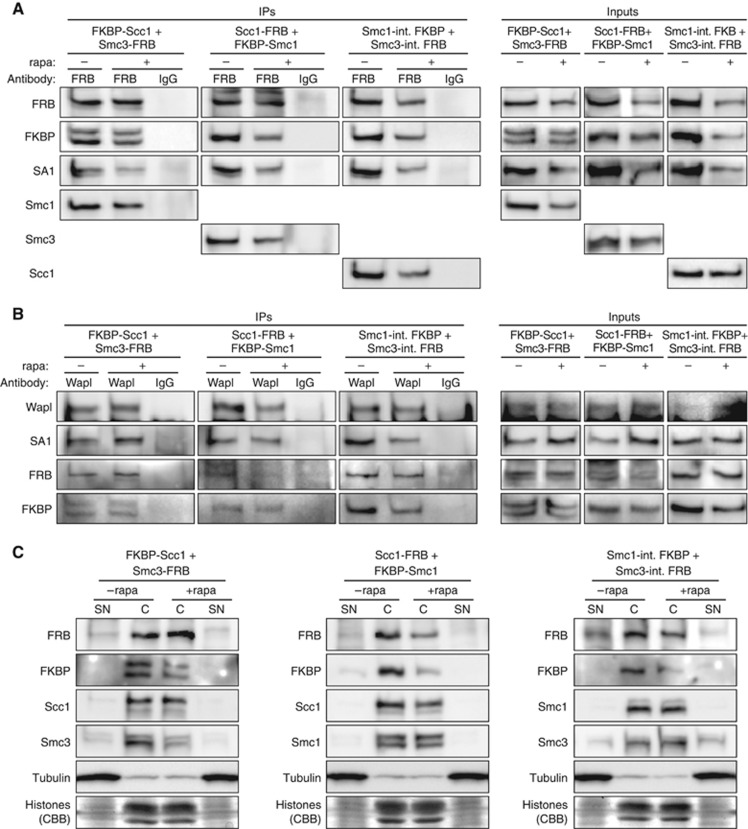
Faithful transmission of chromosomes during eukaryotic cell division requires sister chromatids to be paired from their generation in S phase until their separation in M phase. Cohesion is mediated by the cohesin complex, whose Smc1, Smc3 and Scc1 subunits form a tripartite ring that entraps both DNA double strands. Whereas centromeric cohesin is removed in late metaphase by Scc1 cleavage, metazoan cohesin at chromosome arms is displaced already in prophase by proteolysis-independent signalling. Which of the three gates is triggered by the prophase pathway to open has remained enigmatic. Here, we show that displacement of human cohesin from early mitotic chromosomes requires dissociation of Smc3 from Scc1 but no opening of the other two gates. In contrast, loading of human cohesin onto chromatin in telophase occurs through the Smc1-Smc3 hinge. We propose that the use of differently regulated gates for loading and release facilitates unidirectionality of DNA's entry into and exit from the cohesin ring.


Conflict of interest statement

The authors declare that they have no conflict of interest.

Figures

Figure 1
Figure 1
Localisation and function of cohesin during mitosis. (A, B) RNAi of the prophase pathway component Wapl results in prolonged chromatin association of cohesin during mitosis. HeLa L cells were transfected with siRNA directed against GL2 (Luciferase, control) (A) or Wapl (B). Two days later, their DNA (Hoechst 33342), Hec1 (mitosis-specific kinetochore marker) and Smc1 were visualised by (immuno-) fluorescence microscopy. Scale bars=10 μm. (C) Wapl depletion causes tight cohesion of sister chromatids along their entire length. Two days after transfection of GL2 or Wapl siRNA, HeLa L cells were enriched in prometaphase by a 15-h nocodazole treatment and then subjected to chromosome spreading. Graph depicts the relative number of spreads displaying a primarily ‘Butterfly-like’ or ‘Zipped’ chromosome morphology as exemplified by the images on the right. n=100.
Figure 2
Figure 2
In vivo expression of tailored cohesins with individually lockable gates. (A) Rapamycin can induce heterodimerisation of FRB/FKBP-tagged proteins at human chromatin. Hek293T cells transiently transfected to co-express histone 2B-FRB (H2B-FRB) and FKBP-mCherry fusions were arrested in prometaphase in the presence or absence of rapamycin (±rapa) before mitotic chromatin was analysed by fluorescence microscopy for co-localisation of mCherry (right panels) with Hoechst 33342-stained DNA (left panels). Scale bar=10 μm. (B) Drawn to scale cartoons of the matched pairs of FRB/FKBP-tagged cohesin subunits used in this study. The lines connecting two boxes represent linker peptides of 9 or 29 amino acids, respectively, in Smc1-int. (internal) FKBP and Smc3-int. FRB or in FKBP-Scc1 and FKBP-Smc1. (C) Rationale of one of the three gates of recombinant cohesin being lockable by rapamycin. As illustrated here for the Smc3–Scc1 contact site, each of the three pairs of tailored cohesin subunits will present the FRB and FKBP tags closely juxtaposed at a given gate of the tripartite ring. The rapamycin-induced tight FRB-FKBP interaction should prevent passage of DNA (not shown) through the gate even if the corresponding cohesin subunits detach from each other in response to prophase pathway signalling. (D) Three double-transgenic cell lines inducibly co-express FKBP-Scc1 and Smc3-FRB, Scc1-FRB and FKBP-Smc1 or Smc1-int. FKBP and Smc3-int. FRB. The corresponding Hek293 lines were cultured in the presence or absence of doxycycline (dox) for 24 h before being analysed by FKBP and FRB immunoblots for transgene expression. An anti-α-tubulin western served as loading control. (E) RNAi and transgene induction result in efficient replacement of endogenous cohesin subunits by their FRB/FKBP-tagged counterparts. The double-transgenic Hek293 lines were transfected with siRNAs targeting GL2 (luciferase, control) or the indicated endogenous cohesin subunits and incubated for 3 days in the presence (+) or absence (−) of transgene-inducing doxycycline. Note that Smc3-FRB, Scc1-FRB and Smc3-int. FRB migrate only slightly above the untagged proteins and, thus, are difficult to discern from the endogenous subunits in the mock-depleted samples. Note also that the western signals for Scc1-FRB and Smc3-FRB do not accurately reflect their expression levels because the corresponding antibodies display a greatly reduced sensitivity when their antigens are C-terminally tagged.
Figure 3
Figure 3
The FRB/FKBP-tagged cohesin subunits incorporate into bona fide cohesin complexes capable of interacting with Wapl and chromatin. (A) FRB-tagged cohesin subunits are proficient in rapamycin-independent association with FKBP-tagged and endogenous cohesin subunits. The double-transgenic Hek293 lines were transfected with siRNAs targeting GL2 (luciferase, control) or the respective pair of cohesin subunits that was being replaced by the engineered variants and incubated for 3 days in the presence of doxycycline. Forty-one hours after siRNA transfection, cells were treated with rapamycin (rapa) or DMSO as control. The cells were arrested in prometaphase by a 16-h nocodazole treatment before they were harvested. Cell lysates were cleared by high-speed centrifugation to remove chromatin (Inputs) and then subjected to immunoprecipitation (IPs) with anti-FRB or unspecific IgG. Inputs and immunoprecipitates were finally analysed by western using the indicated antibodies. (B) The essential prophase pathway factor Wapl interacts with FRB/FKBP-tagged cohesin subunits irrespective of the presence or absence of rapamycin. The experiment was conducted as described in (A) with the exception that anti-Wapl was used for the IP instead of anti-FRB. (C) The FRB/FKBP-tagged cohesin subunits retain their ability to bind chromatin. The double-transgenic Hek293 lines were largely treated as described in (A) but without the nocodazole-mediated synchronisation. Cell lysates were prepared in the presence of nuclei permeabilising detergent and centrifuged to separate soluble supernatant (SN) from pelleting chromatin (C), both of which were subsequently analysed by western blot using the specified antibodies and by staining with Coomassie Brilliant Blue (CBB) to visualise histones.
Figure 4
Figure 4
DNA exits the cohesin ring through the Smc3–Scc1 gate. (A) Assay timeline for results shown in (BF). (B) Indicated double-transgenic cell lines were treated as illustrated in (A) and then subjected to chromosome spreading and quantitative assessment of arm cohesion. The relative numbers of cells displaying zipped prometaphase chromosomes are plotted. Each column represents the mean of three independent data points (represented by circles, triangles and squares) totalling 600 analysed cells. Corresponding data sets across all cell lines and conditions are identified by identical shapes. (C, D) Indicated double-transgenic cell lines were treated as illustrated in (A) and then Hoechst 33342- and immunostained to in situ-visualise DNA, Hec1 and Smc1 (C) or FRB (D). Exemplary images are shown. Scale bars=10 μm. (E, F) Quantification of the data shown in (C) and (D), respectively. The relative numbers of cells with prometaphase chromatin positive for Smc1 (E) or FRB (F) are plotted. Circles, triangles and squares correspond to individual data points of three independent reiterations while columns represent means. Data sets of each experiment across all cell lines and conditions are represented by identical shapes. Between 293 and 600 cells were analysed per column.
Figure 5
Figure 5
DNA does not leave cohesin through the Scc1–Smc1 gate. (A) A stable transgenic Hek293 cell line that inducibly expresses a covalent Scc1–Smc1 fusion. The siRNA-resistant expression of the Scc1–Smc1 fusion protein with respect to the endogenous Smc1 that persists after 2 days of RNAi is shown. (B) Covalent fusion of Scc1 and Smc1 is compatible with chromatin association. Insoluble chromatin (C) and soluble supernatant (SN) fractions of the Scc1–Smc1 expressing cell line were analysed by Smc1- and α-tubulin-westerns and by Coomassie Brilliant Blue (CBB) staining of chromatin bound histones. (C, D) The Scc1–Smc1 fusion protein interacts with endogenous cohesin subunits and Wapl. The transgenic Hek293 line was largely treated as described in Figure 3A (only without addition of rapamycin). The respective mitotic cell lysate (Input) was cleared by high-speed centrifugation to remove chromatin and subjected to immunoprecipitations (IPs) with anti-Smc3 (C) or anti-Wapl (D). Co-purifying proteins were detected by western analysis using the indicated antibodies. Mock-IPs using unspecific rabbit IgG served as controls. (E, F) A covalent Scc1–Smc1 fusion does not impair release of cohesin from prophase chromosomes. Scc1–Smc1 expressing cells were treated largely as described in Figure 4A (only without addition of rapamycin) and then analysed by IF (E) and chromosome spreading (F). (E) Averaged over two independent experiments, 99.5% of a total of 200 Hoechst 33342, anti-Hec1, and anti-Smc1 stained cells did not exhibit any signs of Smc1–chromatin association beyond prophase. (F) Averaged over three independent experiments, 94.7% of a total of 300 spread nuclei displayed butterfly-like chromosomes. An increase in tightly cohesed (‘zipped’) chromosomes could not be detected.
Figure 6
Figure 6
Cohesin loading requires opening of the Smc1–Smc3 hinge. (A, B) A double-transgenic cell line induced to co-express Smc1-int. FKBP and Smc3-int. FRB was simultaneously depleted of endogenous Smc1 and -3 by transfection of corresponding siRNAs, nocodazole arrested in prometaphase and finally released into telophase/early G1 phase in the presence or absence of rapamycin (±rapa). (A) Following pre-extraction, fixation and staining of DNA and FRB, only those cells were analysed that were still in telophase or formed closely coupled pairs with similar FRB levels. Exemplary images are shown. Scale bar=10 μm. (B) Quantitative assessment of recombinant cohesin loading. The relative numbers of cell pairs exhibiting FRB staining in telo-/early G1 phase are plotted. Columns correspond to means of three independent experiments with each triplicate totalling 600 cells. Circles, triangles and squares represent data points and identify respective sets from individual experiments. (C, D) Cells were treated as described in (A, B) and stained for FRB and endogenous Scc1. Most of the early G1-phase nuclei that lacked an FRB signal could still reload endogenous Scc1 in the presence of Smc1-int. FKBP, Smc3-int. FRB and rapamycin (due to incomplete knockdown of endogenous Smc1/3 and the rapamycin-independent loading of cohesin complexes that retain at least one untagged SMC subunit). However, the intensities of the Scc1 signals were reduced consistent with the transgenic proteins exerting a dominant-negative effect on cohesin reloading (see text for details). Scale bar=10 μm. For each quantification, 100 cells were counted.
Figure 7
Figure 7
Model of the dynamic cohesin–chromosome interactions throughout the metazoan cell cycle. DNA enters the tripartite cohesin ring through the Smc1–Smc3 gate in telophase. Cohesion is then established during DNA replication in S phase. The bulk of cohesin is released from chromosome arms in prophase of mitosis when DNA exits the ring through the Smc3–Scc1 gate. (Note that sister chromatid separation occurs only when prevailing centromeric cohesin is proteolytically cleaved by separase at the metaphase-to-anaphase transition. This step was omitted for sake of clarity.)

References

    1. Anderson DE, Losada A, Erickson HP, Hirano T (2002) Condensin and cohesin display different arm conformations with characteristic hinge angles. J Cell Biol 156: 419–424 - PMC - PubMed
    1. Banaszynski LA, Liu CW, Wandless TJ (2005) Characterization of the FKBP.rapamycin.FRB ternary complex. J Am Chem Soc 127: 4715–4721 - PubMed
    1. Boos D, Kuffer C, Lenobel R, Körner R, Stemmann O (2008) Phosphorylation-dependent binding of cyclin B1 to a Cdc6-like domain of human separase. J Biol Chem 283: 816–823 - PubMed
    1. Chan K-L, Roig MB, Hu B, Beckouët F, Metson J, Nasmyth K (2012) Cohesin's DNA exit gate is distinct from its entrance gate and is regulated by acetylation. Cell 150: 961–974 - PMC - PubMed
    1. Ciosk R, Shirayama M, Shevchenko A, Tanaka T, Tóth A, Shevchenko A, Nasmyth K (2000) Cohesin's binding to chromosomes depends on a separate complex consisting of Scc2 and Scc4 proteins. Mol Cell 5: 243–254 - PubMed

Publication types

MeSH terms

LinkOut - more resources