Review
. 2013 Apr 21;42(8):3441-52.
doi: 10.1039/c3cs35458j. Epub 2013 Jan 29.

Engineering of bacterial phytochromes for near-infrared imaging, sensing, and light-control in mammals

Affiliations
Review

Engineering of bacterial phytochromes for near-infrared imaging, sensing, and light-control in mammals

Kiryl D Piatkevich et al. Chem Soc Rev. .

Abstract

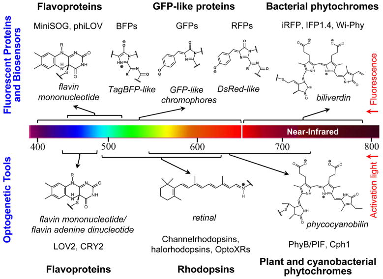
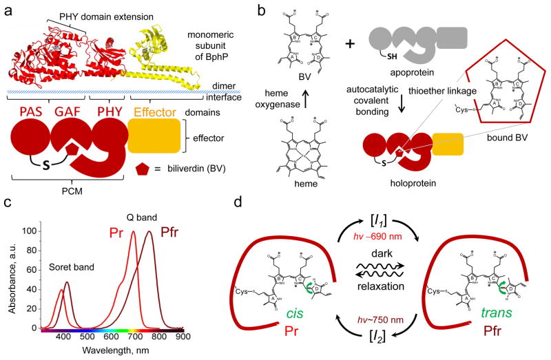
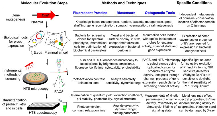
Near-infrared light is favourable for imaging in mammalian tissues due to low absorbance of hemoglobin, melanin, and water. Therefore, fluorescent proteins, biosensors and optogenetic constructs for optimal imaging, optical readout and light manipulation in mammals should have fluorescence and action spectra within the near-infrared window. Interestingly, natural Bacterial Phytochrome Photoreceptors (BphPs) utilize the low molecular weight biliverdin, found in most mammalian tissues, as a photoreactive chromophore. Due to their near-infrared absorbance BphPs are preferred templates for designing optical molecular tools for applications in mammals. Moreover, BphPs spectrally complement existing genetically-encoded probes. Several BphPs were already developed into the near-infrared fluorescent variants. Based on the analysis of the photochemistry and structure of BphPs we suggest a variety of possible BphP-based fluorescent proteins, biosensors, and optogenetic tools. Putative design strategies and experimental considerations for such probes are discussed.


Figures

Fig. 1
Fig. 1
A diversity of the chromophores in the major groups of currently available fluorescent proteins, fluorescent biosensors, and optogenetic tools developed for biotechnological applications are shown. The upper part of the figure shows the chemical structures of flavin mononucleotide, TagBFP-like, GFP-like, DsRed-like and biliverdin chromophores for the respective fluorescent proteins and biosensors derived from flavoproteins (MiniSOG, phiLOV), GFP-like proteins (BFPs, GFPs, RFPs), , and bacterial phytochromes (iRFP, IFP1.4, Wi-Phy). The lower part of the figure shows the chemical structures of flavin mononucleotide, retinal and phycocyanobilin chromophores for the respective optogenetic tools derived from flavoproteins (LOV2, CRY2), rhodopsins (channelrhodopsins, halorhodopsisns, OptoXRs), plant and cyanobacterial phytochromes (PhyB/PIF, Cph1). The chromophores are shown in their protein-linked forms. A color scale presents the wavelength range of fluorescence emission for the fluorescent proteins and biosensors, and the wavelength range of the activation/de-activation light for the optogenetic tools.
Fig. 2
Fig. 2
Structure, formation, spectral and photochemical properties of bacterial phytochromes. (a) Structural organization of a monomer subunit of BphP, (b) synthesis of biliverdin IXα (BV) from heme and its incorporation by apoprotein, (c) absorbance spectra of BphPs in the Pr and Pfr states, and (d) photocycle of BV chromophore within the protein environment are shown. (a, top) Structure of the monomer subunit of the BphP photosensory module (PMC) of Pseudomonas aeruginosa in red (PDB accession ID 3C2W) is overlapped with the structure of the effector domain, represented by histidine kinase in yellow (PDB accession ID 2C2A). (a, bottom) Schematic representation of BphP consisting of the PAS, GAF, PHY, and effector domains. A PHY domain’s extension shields BV from solvent and plays a role in BphP photoconversion. Dimer interface is formed by α-helices of the GAF domain and linker between PMC and effector domain. (b) Degradation of heme to BV is catalyzed by heme oxygenase. This reaction proceeds through a common mechanism that leads to formation of BV, which then autocatalytically covalently attaches to conservative Cys residue in the PAS domain of an apoprotein via a thioether linkage, resulting in a haloprotein. (c) Absorbance spectra of the typical Pr and Pfr states presenting the Q and Soret absorbance bands. (d) BV chromophre in the Pr and Pfr states is shown within protein environment of BphP (dark red curve). Transition from the Pr state to the Pfr state and vice versa is induced with 690 nm and 750 nm light, respectively. The transitions result from rotation of D-ring of the BV chromophore around adjacent double bond (green arrow). In the dark the photoconverted state undergoes spontaneous relaxation back to the ground state (waved arrows). The transition from the Pr to Pfr state and vice versa occurs via different intermediate states I1 and I2, respectively.
Fig. 3
Fig. 3
Alignment of amino acid sequences of the photosensory modules of the most characterized BphPs. The proteins were chosen based on the availability of the crystal structures (PaBphP, RpBphP3, DrBphP) and those that were developed to the fluorescent proteins (IFP1.4, iRFP, Wi-Phy, and RpBphP2 as the template for iRFP). The numbering of amino acid residues follows that for the PaBphP protein. Cys residue, which is covalently attached to the BV chromophore, is marked with asterisk. The chromophore surrounding residues within 4.5 Å, 4.5–5.5 Å and 5.5–6.5 Å are highlighted with gray, cyan, and red colors, respectively. The residues located in the dimer interface are highlighted with yellow. The residues located in the close proximity to the thioether bond between BV and apoprotein are underlined. The α-helixes and β-sheets demonstrate the secondary structure of BphPs. The PAS, GAF and PHY domains are underlined with the blue, green, and red lines, respectively.
Fig. 4
Fig. 4
Proposed genetically-encoded near-infrared (NIR) probes based on bacterial phytochromes: (a) versatile two-domain short-NIR and three-domain long-NIR fluorescent proteins (FPs), photoactivatable (PA) and photoswitchable (PS) three-domain NIR fluorescent proteins, (b) two-domain biosensors for redox status and metal ions (Men+), split biosensors for protein interactions resulted from enzymatic modifications, such as phosphorylation (designated as P−), and insertion-based biosensors to detect analytes, and (c) optogenetic tools controlling enzymatic activities, open and closed states of ion channels, and gene expression via regulation of interaction between DNA repressor and gene promoter. The schematic illustration of the structural elements of BphPs corresponds to those shown in Figure 2a. Please see text for more details.
Fig. 5
Fig. 5
Molecular evolution steps, methods and techniques, and specific conditions in the course of development of the BphP-based NIR fluorescent proteins, biosensors, and optogenetic tools. Vertical arrows indicate the typical order of the evolution steps such as gene construction and mutagenesis, biological hosts for protein expression, instrumental methods of screening, protein characterization in vitro and in cells. Methods and techniques proposed for each molecular evolution step are subdivided per the proposed NIR probes. Specific conditions indicate particular qualities of BphPs that should be considered for each directed evolution step. HTS is a high-throughput screening, FACS is a fluorescence-activated cell sorter, and λ is a wavelength. See also Table 2 for details on knowledge-based mutagenesis.

References

    1. van der Horst MA, Hellingwerf KJ. Acc Chem Res. 2004;37:13–20. - PubMed
    1. Wu B, Piatkevich KD, Lionnet T, Singer RH, Verkhusha VV. Curr Opin Cell Biol. 2011;23:310–317. - PMC - PubMed
    1. Subach FV, Verkhusha VV. Chem Rev. 2012;112:4308–4327. - PMC - PubMed
    1. Piatkevich KD, Verkhusha VV. Curr Opin Chem Biol. 2010;14:23–29. - PMC - PubMed
    1. Gomelsky M, Hoff WD. Trends Microbiol. 2011;19:441–448. - PubMed

Publication types