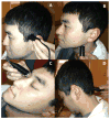
Transcranial Doppler ultrasound: technique and application
- PMID: 23361485
- PMCID: PMC3902805
- DOI: 10.1055/s-0032-1331812
Transcranial Doppler ultrasound: technique and application
Abstract
Transcranial Doppler (TCD) ultrasound provides rapid, noninvasive, real-time measures of cerebrovascular function. TCD can be used to measure flow velocity in the basal arteries of the brain to assess relative changes in flow, diagnose focal vascular stenosis, or to detect embolic signals within these arteries. TCD can also be used to assess the physiologic health of a particular vascular territory by measuring blood flow responses to changes in blood pressure (cerebral autoregulation), changes in end-tidal CO2 (cerebral vasoreactivity), or cognitive and motor activation (neurovascular coupling or functional hyperemia). TCD has established utility in the clinical diagnosis of a number of cerebrovascular disorders such as acute ischemic stroke, vasospasm, subarachnoid hemorrhage, sickle cell disease, as well as other conditions such as brain death. Clinical indication and research applications for this mode of imaging continue to expand. In this review, the authors summarize the basic principles and clinical utility of TCD and provide an overview of a few TCD research applications.
Thieme Medical Publishers 333 Seventh Avenue, New York, NY 10001, USA.
Figures


Similar articles
-
Applications of transcranial Doppler in the ICU: a review.Intensive Care Med. 2006 Jul;32(7):981-94. doi: 10.1007/s00134-006-0173-y. Epub 2006 May 10. Intensive Care Med. 2006. PMID: 16791661 Review.
-
Transcranial Doppler: a stethoscope for the brain-neurocritical care use.J Neurosci Res. 2018 Apr;96(4):720-730. doi: 10.1002/jnr.24148. Epub 2017 Sep 7. J Neurosci Res. 2018. PMID: 28880397 Review.
-
Transcranial Doppler ultrasonography in neurological surgery and neurocritical care.Neurosurg Focus. 2019 Dec 1;47(6):E2. doi: 10.3171/2019.9.FOCUS19611. Neurosurg Focus. 2019. PMID: 31786564 Review.
-
Transcranial Doppler and Optic Nerve Sonography.J Cardiothorac Vasc Anesth. 2019 Aug;33 Suppl 1:S38-S52. doi: 10.1053/j.jvca.2019.03.040. J Cardiothorac Vasc Anesth. 2019. PMID: 31279352 Review.
-
Utility of transcranial Doppler ultrasound for the integrative assessment of cerebrovascular function.J Neurosci Methods. 2011 Mar 30;196(2):221-37. doi: 10.1016/j.jneumeth.2011.01.011. Epub 2011 Jan 27. J Neurosci Methods. 2011. PMID: 21276818 Review.
Cited by
-
Bridging the Divide: Brain and Behavior in Developmental Language Disorder.Brain Sci. 2023 Nov 19;13(11):1606. doi: 10.3390/brainsci13111606. Brain Sci. 2023. PMID: 38002565 Free PMC article. Review.
-
Transcranial Color Coded Duplex Sonography Findings in Stroke Patients Undergoing Rehabilitation: An Observational Study.J Neurosci Rural Pract. 2022 Jan 12;13(1):129-133. doi: 10.1055/s-0041-1742158. eCollection 2022 Jan. J Neurosci Rural Pract. 2022. PMID: 35110933 Free PMC article.
-
Optimization of time domain diffuse correlation spectroscopy parameters for measuring brain blood flow.Neurophotonics. 2021 Jul;8(3):035005. doi: 10.1117/1.NPh.8.3.035005. Epub 2021 Aug 12. Neurophotonics. 2021. PMID: 34395719 Free PMC article.
-
Changes in arterial cerebral blood volume during lower body negative pressure measured with MRI.Neuroimage. 2019 Feb 15;187:166-175. doi: 10.1016/j.neuroimage.2017.06.041. Epub 2017 Jun 28. Neuroimage. 2019. PMID: 28668343 Free PMC article.
-
Neuroinflammation and Precision Medicine in Pediatric Neurocritical Care: Multi-Modal Monitoring of Immunometabolic Dysfunction.Int J Mol Sci. 2020 Dec 1;21(23):9155. doi: 10.3390/ijms21239155. Int J Mol Sci. 2020. PMID: 33271778 Free PMC article. Review.
References
-
- Aaslid R. Transcranial Doppler Sonography. New York: Springer-Verlag; 1986.
-
- DeWitt LD, Wechsler LR. Transcranial Doppler. Stroke. 1988;19(7):915–921. - PubMed
-
- Tegeler CH, Ratanakorn D. Physics and principles. In: Babikian VL, Wechsler LR, editors. Transcranial Doppler Ultrasonography. 2. Waltham, MA: Butterworth-Heinemann; 1999. pp. 3–11.
-
- Bishop CC, Powell S, Rutt D, Browse NL. Transcranial Doppler measurement of middle cerebral artery blood flow velocity: a validation study. Stroke. 1986;17(5):913–915. - PubMed
-
- Nuttall GA, Cook DJ, Fulgham JR, Oliver WC, Jr, Proper JA. The relationship between cerebral blood flow and transcranial Doppler blood flow velocity during hypothermic cardiopulmonary bypass in adults. Anesth Analg. 1996;82(6):1146–1151. - PubMed
Publication types
MeSH terms
Grants and funding
LinkOut - more resources
Full Text Sources
Other Literature Sources
Medical

