

CT imaging features of eosinophilic colitis in children
- PMID: 23361493
- PMCID: PMC3816107
- DOI: 10.1007/s00247-012-2615-8
CT imaging features of eosinophilic colitis in children
Abstract
Background: Eosinophilic colitis (EC) is a gastrointestinal disease of undetermined etiology whose clinical features overlap with those of the inflammatory bowel diseases. To the best of our knowledge, the CT imaging features of EC have not been described in children.
Objective: To report and analyze the clinical, imaging and histological findings in seven children with EC.
Materials and methods: Children with EC were identified in a pediatric pathology database, and those with CT imaging within 2 months of diagnosis were included, totaling seven children. Clinical, imaging and pathological features were reviewed and analyzed.
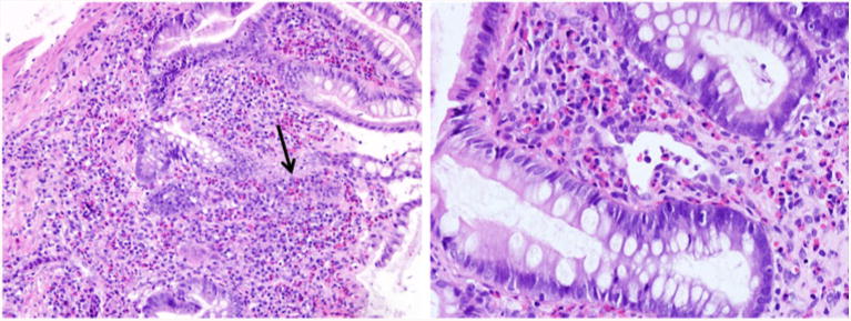
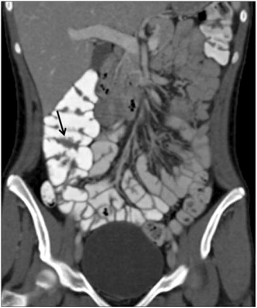
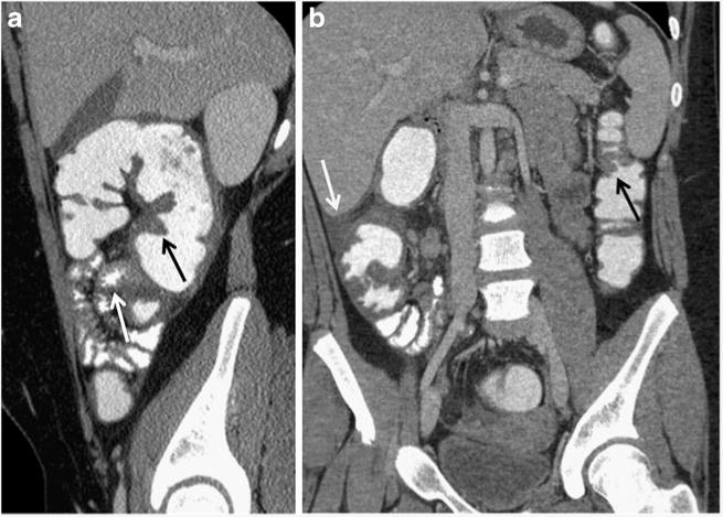
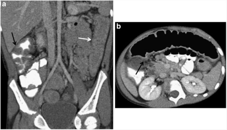
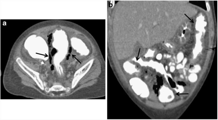
Results: The most common presenting symptoms were abdominal pain, bloody diarrhea and rectal bleeding. EC was characterized as a dense and predominantly eosinophilic inflammatory infiltrate in the lamina propria or epithelium without granulomas. CT scans were abnormal in six children (86%), demonstrating colonic wall thickening, predominantly cecal, in five (71%), mild to moderate terminal ileal thickening in two (29%), and pneumatosis in one (14%). Right colonic involvement was greater than terminal ileal involvement.
Conclusion: CT imaging findings in children with EC include right colonic wall thickening of variable extent downstream and absent or mild involvement of the terminal ileum. EC should be considered in the differential diagnosis in children presenting with abdominal pain and bloody diarrhea.
Conflict of interest statement
Figures
References
-
- Rothenberg ME. Eosinophilic gastrointestinal disorders (EGID) J Allergy Clin Immunol. 2004;113:11–28. - PubMed
-
- Klein NC, Hargrove RL, Sleisenger MH, et al. Eosinophilic gastroenteritis. Medicine (Baltimore) 1970;49:299–319. - PubMed
-
- Clouse RE, Alpers DH, Hockenbery DM, et al. Pericrypt eosinophilic enterocolitis and chronic diarrhea. Gastroenterology. 1992;103:168–176. - PubMed
-
- Velchuru VR, Khan MA, Hellquist HB, et al. Eosinophilic colitis. J Gastrointest Surg. 2007;11:1373–1375. - PubMed
MeSH terms
Supplementary concepts
Grants and funding
LinkOut - more resources
Full Text Sources
Other Literature Sources
Medical
