

Heterotopic bone formation about the hip undergoes endochondral ossification: a rabbit model
- PMID: 23361932
- PMCID: PMC3613540
- DOI: 10.1007/s11999-013-2801-5
Heterotopic bone formation about the hip undergoes endochondral ossification: a rabbit model
Abstract
Background: Heterotopic ossification (HO) occurs most commonly after trauma and surgery about the hip and may compromise subsequent function. Currently available animal models describing the cellular progression of HO are based on exogenous osteogenic induction agents and may not reflect the processes following trauma.
Questions/purposes: We therefore sought to characterize the histologic progression of heterotopic bone formation in an animal model that recapitulates the human condition without the addition of exogenous osteogenic material.
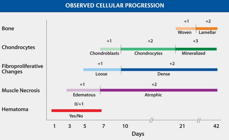
Methods: We used a rabbit model that included intramedullary instrumentation of the upper femur and ischemic crush injury of the gluteal muscle. Bilateral surgical induction procedures were performed on 30 animals with the intention of inciting the process of HO; no supplemental osteogenic stimulants were used. Three animals were sacrificed at each of 10 predetermined times between 1 day and 26 weeks postoperatively and the progression of tissue maturation was graded histologically using a five-item scale.
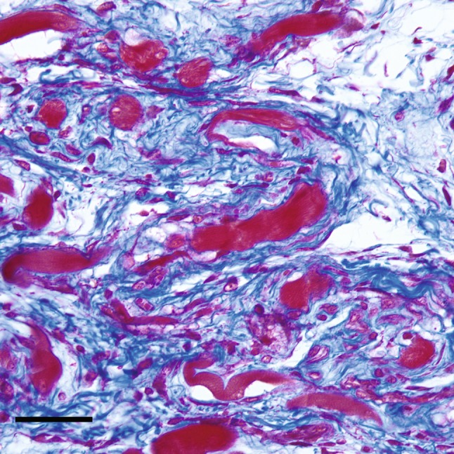
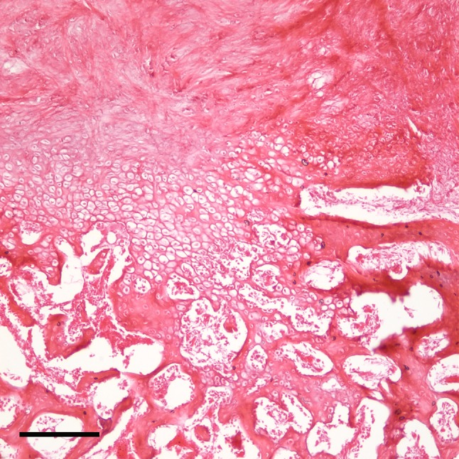
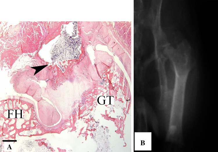
Results: Heterotopic bone reliably formed de novo and consistently followed a pathway of endochondral ossification. Chondroid elements were found in juxtaposition with immature woven bone in all sections that contained mature osseous elements.
Conclusions: These results establish that HO occurs in an animal model mimicking the human condition following surgical trauma about the hip; it is predictable in its histologic progression and follows a pathway of endochondral bone formation.
Clinical relevance: By showing a consistent pathway of endochondral ossification leading to ectopic bone formation, this study provides a basis for understanding the mechanisms by which HO might be mitigated by interventions.
Figures


References
-
- Ahrengart L. Periarticular heterotopic ossification after total hip arthroplasty: risk factors and consequences. Clin Orthop Relat Res. 1991;263:49–58. - PubMed
-
- Ayers DC, Evarts CM, Parkinson JR. The prevention of heterotopic ossification in high-risk patients by low-dose radiation therapy after total hip arthroplasty. J Bone Joint Surg Am. 1986;68:1423–1430. - PubMed
-
- Ayers DC, Pellegrini VD, Jr, Evarts CM. Prevention of heterotopic ossification in high-risk patients by radiation therapy. Clin Orthop Relat Res. 1991;263:87–93. - PubMed
MeSH terms
LinkOut - more resources
Full Text Sources
Other Literature Sources
