

Adult multifocal pigmented villonodular synovitis--clinical review
- PMID: 23361936
- PMCID: PMC3609970
- DOI: 10.1007/s00264-013-1789-5
Adult multifocal pigmented villonodular synovitis--clinical review
Abstract
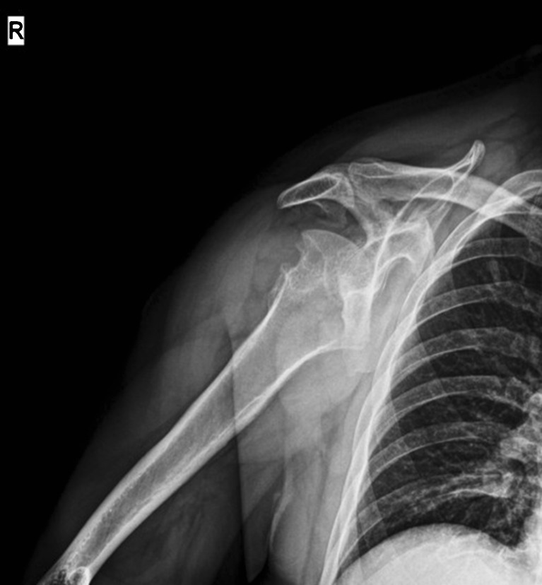
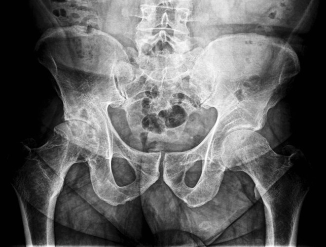
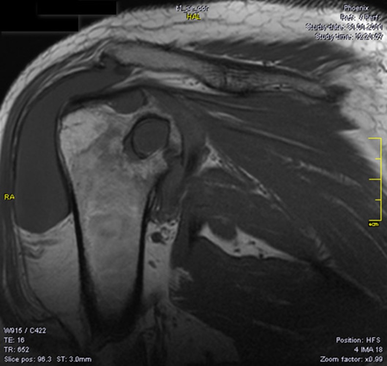
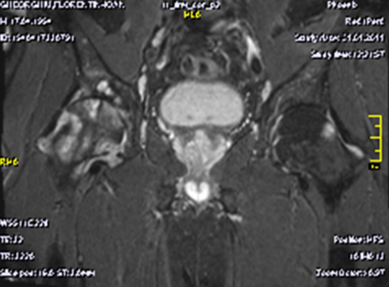
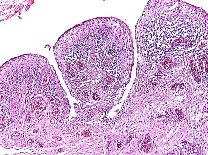
Pigmented villonodular synovitis (PVNS) is a rare, benign proliferative disease of the synovial tissue that affects a single joint or a tendon sheath. Data from the literature present only a few cases of multifocal PVNS. This paper presents multifocal PVNS in the adult. This disease can affect bilateral shoulders, hips and knees. The diagnosis may be delayed by the slow evolution of the disease (up to ten years); some patients may be seen with late-stage degenerative joints, serious complications, painful and functionally uncompensated, with significant locomotion deficit. PVNS requires a radical treatment with prosthetic arthroplasty associated with synovectomy. Complex imaging (X-Rays, magnetic resonance imaging (MRI), ultrasound) and macroscopic appearance of the lesions during surgery confirms the clinical diagnosis of multifocal PVNS with secondary bone lesions. Histology marks the final diagnosis of multifocal PVNS. The postoperative results are good, with recovery in functional parameters of the joints with endoprosthesis.
Figures
References
-
- Byers PD, Cotton RE, Deacon UW. The diagnosis and treatment of pigmented villonodular synovitis. J Bone Joint Surg Am. 1968;50:290–305. - PubMed
-
- Smith JH, Pugh DG. Roentgenographic aspects of articular pigmented villonodular synovitis. AJR Am J Roentgenol. 1962;87:1146–1156. - PubMed
-
- Jaffe HL, Lichtenstein L, Sutro CJ. Pigmented villonodular synovitis, bursitis, and tenosynovitis. Arch Pathol. 1941;31:731–765.
Publication types
MeSH terms
LinkOut - more resources
Full Text Sources
Other Literature Sources
