. 2013 Jan;25(1):257-69.
doi: 10.1105/tpc.112.107045. Epub 2013 Jan 29.

HDA18 affects cell fate in Arabidopsis root epidermis via histone acetylation at four kinase genes

Affiliations

HDA18 affects cell fate in Arabidopsis root epidermis via histone acetylation at four kinase genes

Cui Liu et al. Plant Cell. 2013 Jan.

Abstract

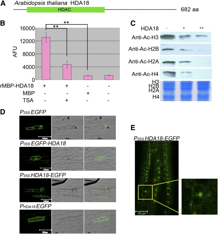
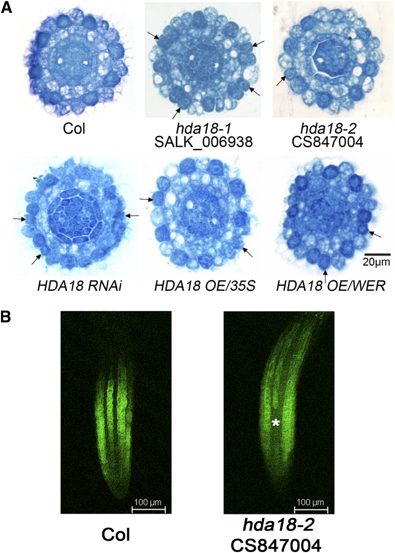
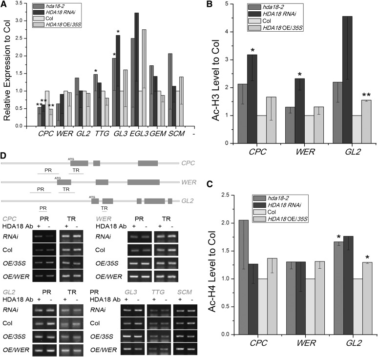
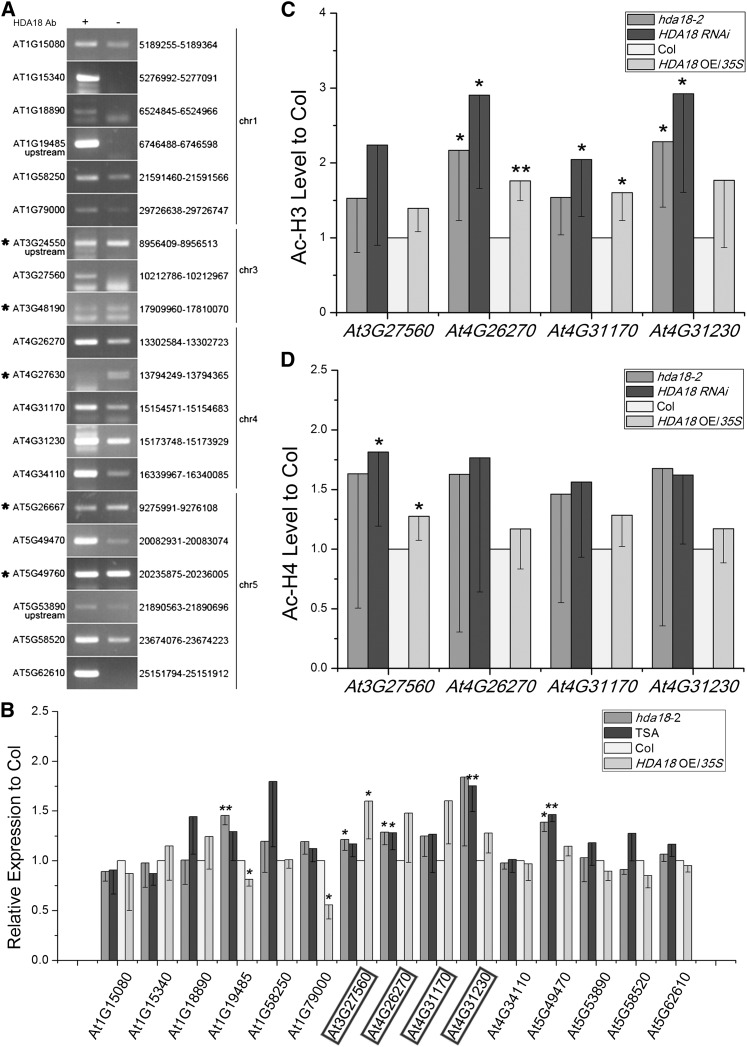
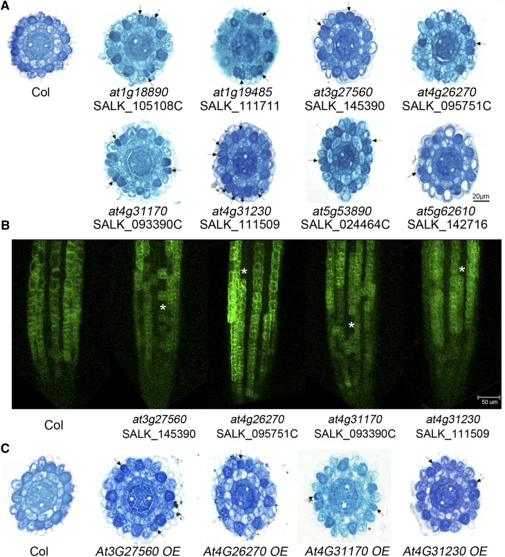
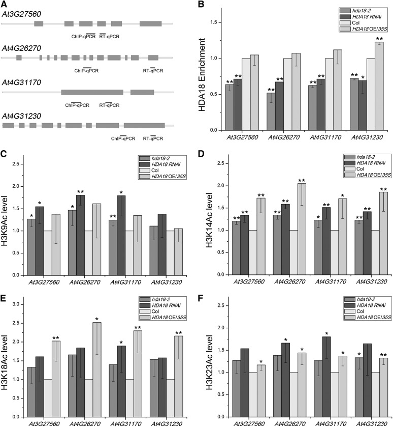
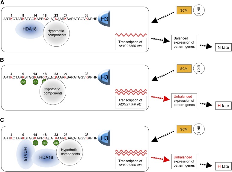
The differentiation of hair (H) and non-hair (N) cells in the Arabidopsis thaliana root epidermis is dependent on positional relationships with underlying cortical cells. We previously found that histone acetylation relays positional information and that a mutant altered in the histone deacetylase gene family member HISTONE DEACETYLASE 18 (HDA18) exhibits altered H and N epidermal cell patterning. Here, we report that HDA18 has in vitro histone deacetylase activity and that both mutation and overexpression of HDA18 led to cells at the N position having H fate. The HDA18 protein physically interacted with histones related to a specific group of kinase genes, which are demonstrated in this study to be components of a positional information relay system. Both down- and upregulation of HDA18 increased transcription of the targeted kinase genes. Interestingly, the acetylation levels of histone 3 lysine 9 (H3K9), histone 3 lysine 14 (H3K14) and histone 3 lysine 18 (H3K18) at the kinase genes were differentially affected by down- or upregulation of HDA18, which explains why the transcription levels of the four HDA18-target kinase genes increased in all lines with altered HDA18 expression. Our results reveal the surprisingly complex mechanism by which HDA18 affects cellular patterning in Arabidopsis root epidermis.


Figures

Figure 1.
Figure 1.
HDA18 Protein Has HDAC Activity and Localizes to Both the Nucleus and Cytoplasm. (A) Schematic of HDA18 protein consisting of 682 amino acids with a conserved HDAC domain (green bar). aa, amino acids. (B) In vitro HDAC activity assay with fluorescent substrate using a recombinant protein consisting of the HDAC domain of HDA18 fused to a maltose binding protein (MBP) tag. The HDAC activity of the recombinant protein was inhibited by TSA, an HDAC-specific inhibitor. Data show mean ± sd from four independent experiments (**P < 0.01; Student’s t test). AFU, arbitrary fluorescence units. (C) In-gel assay revealing that the HDAC domain of HDA18 has similar deacetylation activity on the four core histones. (D) Transient expression of HDA18 protein in onion epidermis revealed that HDA18 protein localizes to both the nucleus and cytoplasm (left column, dark field with florescence of GFP; middle column, bright field; right column, merged). (E) Observation of EGFP localization in a T2 transgenic Arabidopsis line. The small image at the right is an enlargement of the squared area in the left image.
Figure 2.
Figure 2.
Altered HDA18 Expression Confers H Fate to Some Cells at the N Position. (A) Cross sections of root tips of the wild type (Columbia [Col], top row, left), mutants (top row, middle and right), RNAi (bottom row, left), and OE lines (bottom row, middle and right) at the ends of the root caps, showing cells with H fate at N positions (arrows). The regular pattern of epidermal cells in the wild type (Col) is that cells reside over the intercellular space between cortical cells (H position) differentiating to H cells were darkly stained, while those directly over cortical cells (N position) differentiating to N cells were less stained. (B) PGL2:GFP expression in roots of mutant (hda18-2) and the wild type (Col). Since GL2 is specifically expressed in N cells, and root cells form continuous files, the florescence of GFP generated by PGL2:GFP presents as continuous files longitudinally (Col). An asterisk indicates the cells at N positions converted to H fate (no GFP signal detected).
Figure 3.
Figure 3.
Representative Pattern Gene Expression Levels and Histone Acetylation Status of CPC, WER, and GL2 in Lines with Altered HDA18 Expression. (A) Expression levels of pattern genes were altered in root tips of hda18-2 mutant, RNAi, and OE lines. Data show mean ± sd from four to seven independent real-time PCR experiments, each with three replicates (*P < 0.05 and **P < 0.01; Student’s t test). Col, Columbia. (B) and (C) Histone acetylation status of H3 and H4, examined by ChIP-PCR with AcH3 and AcH4 antibodies, at CPC, WER, and GL2 sequences in root tips of hda18-2 mutant, RNAi, and OE lines. Data show mean ± sd from three independent experiments, each with three replicates (*P < 0.05 and **P < 0.01; Student’s t test). (D) HDA18 protein does not bind pattern genes based on ChIP-PCR analysis of regions shown in orange. PR, promoter region; TR, transcribed region.
Figure 4.
Figure 4.
HDA18 Protein Binds and Affects Transcription of Certain Kinase Genes by Changing Their Histone Acetylation Status. (A) ChIP-PCR verification of binding of HDA18 protein to putative targets identified by ChIP-chip. The genes marked with asterisks were false positives. The numbers listed to the right indicate the sites identified in the ChIP-chip analysis (for detailed information, see Supplemental Data Set 1 online). (B) Expression levels of HDA18-targeted genes in root tips with altered HDA18 expression, showing upregulation of four kinase genes (highlighted with frames) regardless of increased or decreased expression of HDA18. Data show mean ± sd from three to four independent experiments, each with three replicates (*P < 0.05 and **P < 0.01; Student’s t test). Col, Columbia. (C) and (D) Histone acetylation status of H3 and H4 correspond to altered HDA18 expression, examined by ChIP-PCR with AcH3 and AcH4 antibodies at the identified binding sites of selected kinase genes. Data show mean ± sd from five independent experiments, each with three replicates (*P < 0.05 and **P < 0.01; Student’s t test).
Figure 5.
Figure 5.
Identification of Kinase Genes as Components Involved in Cellular Patterning of the Root Epidermis. (A) Cross sections of root tips of mutants of verified HDA18 target genes at the ends of root caps, showing cells with H fate at N positions (arrows). Col, Columbia. (B) PGL2:GFP expression in roots of mutants (at3g27560, at4g26270, at4g31170, and at4g31230) and the wild type (Col). Asterisks represent the cells at N positions converted to H fate (no GFP signal detected). (C) Cross sections of root tips of OE lines of selected HDA18 target kinase genes at the ends of root caps, showing cells with H fate at N positions (arrows).
Figure 6.
Figure 6.
Effects of HDA18 Levels on the Acetylation of H3K9, H3K14, H3K18, and H3K23 at the Four HDA18-Target Kinase Genes. (A) Regions selected for ChIP-PCR analyses are indicated with short black bars. qPCR, quantitative PCR. (B) The enrichment of DNA related to the four kinase genes in ChIP assays using HDA18 protein in the wild type and altered HDA18 expression lines. Data show mean ± sd from three independent experiments, each with three replicates (*P < 0.05 and **P < 0.01; Student’s t test). Col, Columbia. (C) to (F) The changes in acetylation of the H3 Lys residues at the four kinase genes under down- and upregulated HDA18 expression examined using antibodies specific to H3K9, H3K14, H3K18, and H3K23. Data show mean ± sd from four independent experiments, each with three replicates (*P < 0.05 and **P < 0.01; Student’s t test).
Figure 7.
Figure 7.
Model of Possible Mechanism for HDA18 Regulation of Transcription of the Four Kinase Genes. The HDA18 target kinase genes function between the membrane-localized receptor-like kinases, such as SCM (orange rectangles) and BRI1 (open circles), and the pattern genes in the GL2-centered TFN (open rectangles). (A) In the wild type, the HDA18 protein, perhaps with other unknown components, binds to the region containing H3K9 through Lys-23 of the H3 related to the four target kinase genes and maintains the acetylation of these residues at the proper levels for balanced transcription of the kinase genes. (B) In the hda18 mutant, the quantity of HDA18 protein is reduced and the region containing H3K9 through K23 of the H3 related to the four kinase genes cannot be properly covered. The acetylation levels are therefore increased (green circles), which results in increased transcription of the four kinase genes (red waves). The latter disturbs the previously balanced expression of the pattern genes and results in the cell conversion phenotype. (C) In the HDA18 OE line, surplus HDA18 protein may interfere with the interaction of HDA18 with the unknown components and lead to an abnormal structure, leaving H3K14 and H3K18 unbound by HDA18. This would result in increased acetylation at those sites, which results in increased transcription of the four kinase genes. The latter disturbs the previously balanced expression of pattern genes and results in the cell conversion phenotype similar to that seen in the hda18 mutants.

Comment in

  • Trust in nature.
    Bai SN. Bai SN. Plant Signal Behav. 2013 May;8(5):e23936. doi: 10.4161/psb.23936. Epub 2013 Feb 20. Plant Signal Behav. 2013. PMID: 23425922 Free PMC article.

References

    1. Alinsug M.V., Yu C.W., Wu K. (2009). Phylogenetic analysis, subcellular localization, and expression patterns of RPD3/HDA1 family histone deacetylases in plants. BMC Plant Biol. 9: 37. - PMC - PubMed
    1. Berger S.L. (2007). The complex language of chromatin regulation during transcription. Nature 447: 407–412 - PubMed
    1. Birnbaum K., Shasha D.E., Wang J.Y., Jung J.W., Lambert G.M., Galbraith D.W., Benfey P.N. (2003). A gene expression map of the Arabidopsis root. Science 302: 1956–1960 - PubMed
    1. Brady S.M., Orlando D.A., Lee J.Y., Wang J.Y., Koch J., Dinneny J.R., Mace D., Ohler U., Benfey P.N. (2007). A high-resolution root spatiotemporal map reveals dominant expression patterns. Science 318: 801–806 - PubMed
    1. Caro E., Castellano M.M., Gutierrez C. (2007). A chromatin link that couples cell division to root epidermis patterning in Arabidopsis. Nature 447: 213–217 - PubMed

Publication types

MeSH terms