. 2013 Jan;4(1):106-17.
doi: 10.18632/oncotarget.805.

Quantification of Mesenchymal Stem Cells (MSCs) at sites of human prostate cancer

Affiliations

Quantification of Mesenchymal Stem Cells (MSCs) at sites of human prostate cancer

W Nathaniel Brennen et al. Oncotarget. 2013 Jan.

Abstract

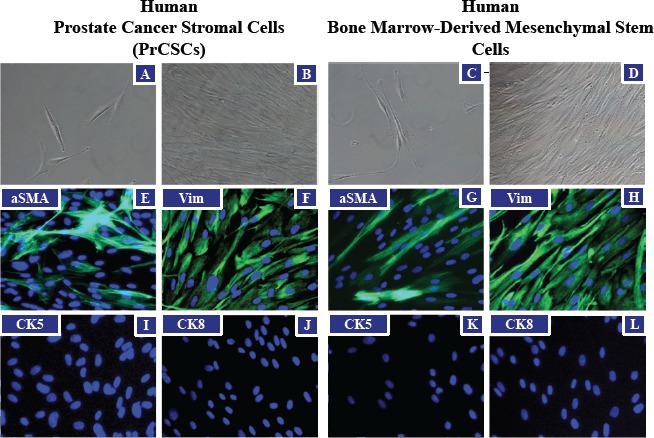
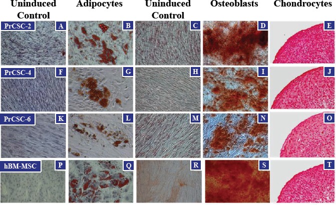
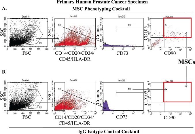
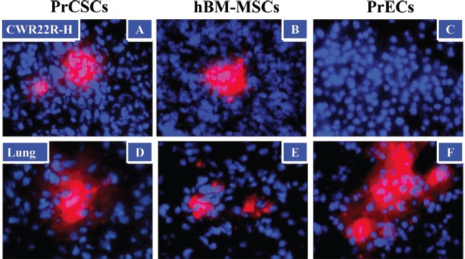
Circulating bone marrow-derived Mesenchymal Stem Cells (BM-MSCs) have an innate tropism for tumor tissue in response to the inflammatory microenvironment present in malignant lesions. The prostate is bombarded by numerous infectious and inflammatory insults over a lifetime. Chronic inflammation is associated with CXCL12, CCL5, and CCL2, which are highly overexpressed in prostate cancer. Among other cell types, these chemoattractant stimuli recruit BM-MSCs to the tumor. MSCs are minimally defined as plastic-adhering cells characterized by the expression of CD90, CD73, and CD105 in the absence of hematopoietic markers, which can differentiate into osteoblasts, chondrocytes, and adipocytes. MSCs are immunoprivileged and have been implicated in tumorigenesis through multiple mechanisms, including promoting proliferation, angiogenesis, and metastasis, in addition to the generation of an immunosuppressive microenvironment. We have demonstrated that MSCs represent 0.01-1.1% of the total cells present in core biopsies from primary human prostatectomies. Importantly, these analyses were performed on samples prior to expansion in tissue culture. MSCs in these prostatectomy samples are FAP-, CD90-, CD73-, and CD105-positive, and CD14-, CD20-, CD34-, CD45-, and HLA-DR-negative. Additionally, like BM-MSCs, these prostate cancer-derived stromal cells (PrCSCs) were shown to differentiate into osteoblasts, adipocytes and chondrocytes. In contrast to primary prostate cancer-derived epithelial cells, fluorescently-labeled PrCSCs and BM-MSCs were both shown to home to CWR22RH prostate cancer xenografts following IV injection. These studies demonstrate that not only are MSCs present in sites of prostate cancer where they may contribute to carcinogenesis, but these cells may also potentially be used to deliver cytotoxic or imaging agents for therapeutic and/or diagnostic purposes.


Figures

Figure 1
Figure 1. Morphological Similarities between PrCSCs and hBM-MSCs
Prostate cancer-derived stromal cells (PrCSCs) and human bone marrow-derived mesenchymal stem cells (hBM-MSCs) have similar morphologies at low (A and C) and high (B and D) densities (representative phase-contrast images). Both PrCSCs and hBM-MSCs stain positive for mesenchymal markers, alpha-smooth muscle actin (aSMA) (green, E and G) and vimentin (Vim) (green, F and H), but not epithelial markers, cytokeratin 5 (I and K) or cytokeratin 8 (J and L) by immunofluorescence. Nuclei counterstained with DAPI (blue, E-L).
Figure 2
Figure 2. Multi-lineage Differentiation of PrCSCs and hBM-MSCs
PrCSCs derived from multiple patients (PrCSC-2, -4, -6) are able to differentiate into adipocytes (B, G, and L), osteoblasts (D, I, and N), and chondrocytes (E, J, and O) when placed in the appropriate induction media as defined by positive staining for lipid vacuoles (adipocytes, Oil Red O), calcium mineralization (osteoblasts, Alizarin Red S), and glycosaminoglycans (chondrocytes, Safranin-O), respectively. Differentiation indicated by red staining in each. In contrast, no differentiation is observed when these cells are not cultured in the presence of the various inducing factors (adipocytes: A, F, K, and P; osteoblasts: C, H, M, and R). Differentiation into these three lineages is one of the defining characteristics of mesenchymal stem cells as demonstrated by the hBM-MSC positive controls (Q, S, and T).
Figure 3
Figure 3. Method for Quantifying MSCs in Primary Human Prostatectomy Samples
MSCs were quantified from primary human prostatectomy specimens using an optimized flow cytometry assay (A-B). Prostatectomy samples were digested into a single cell suspension using a combination of enzymatic and mechanical methods. At least 10,000 cells were initially gated (R1) on the basis forward and side scatter (FSC and SSC, respectively). From this initial population, lineage-negative cells (CD14, CD20, CD34, CD45, HLA-DR) were selected (R2) and analyzed for expression of CD73 (R3). These lineage-negative, CD73-positive cells were further analyzed for the co-expression of CD90 and CD105. MSCs were defined as being lineage-negative and triple-positive for CD73, CD90, and CD105 (red box). Final quantification was performed by subtracting the number of events meeting these criteria in the IgG isotype control cocktail analysis (red box, B) from the events detected in the sample stained with the MSC phenotyping cocktail (red box, A). Importantly, all samples were analyzed within 3 hrs post-surgery.
Figure 4
Figure 4. Tumor Trafficking of PrCSCs and hBM-MSCs to Human Cancer Xenografts in Mice
PrCSCs (A) and hBM-MSCs (B), but not PrECs (C), traffic to prostate cancer xenografts in vivo following systemic infusion. Fluorescently-labeled (CM-DiI, red) PrCSCs, hBM-MSCs, and PrECs (1 × 10⁶) were infused intravenously (IV) into immunocompromised mice bearing subcutaneous CWR22RH xenografts (3/group). Four days post-infusion, lungs and tumors were harvested and analyzed by fluorescence microscopy for the presence of CM-DiI-labeled cells. In contrast to the xenografts, all three cell types were found entrapped in the lungs following infusion (D-F). Nuclei counterstained with DAPI (blue). At least three images analyzed per tissue per animal, representative images shown.

References

    1. De Marzo AM, Platz EA, Sutcliffe S, Xu J, Gronberg H, Drake CG, Nakai Y, Isaacs WB, Nelson WG. Inflammation in prostate carcinogenesis. Nat Rev Cancer. 2007;7(4):256–269. - PMC - PubMed
    1. Sfanos KS, De Marzo AM. Prostate cancer and inflammation: the evidence. Histopathology. 2012;60(1):199–215. - PMC - PubMed
    1. English K, Mahon BP. Allogeneic mesenchymal stem cells: agents of immune modulation. J Cell Biochem. 2011;112(8):1963–1968. - PubMed
    1. Caplan AI. Why are MSCs therapeutic? New data: new insight. J Pathol. 2009;217(2):318–324. - PMC - PubMed
    1. Bianchi G, Borgonovo G, Pistoia V, Raffaghello L. Immunosuppressive cells and tumour microenvironment: focus on mesenchymal stem cells and myeloid derived suppressor cells. Histol Histopathol. 2011;26(7):941–951. - PubMed

Publication types

MeSH terms