. 2013 Mar 29;288(13):8815-25.
doi: 10.1074/jbc.M112.440503. Epub 2013 Jan 29.

Mechanisms governing the endosomal membrane recruitment of the core retromer in Arabidopsis

Affiliations

Mechanisms governing the endosomal membrane recruitment of the core retromer in Arabidopsis

Enric Zelazny et al. J Biol Chem. .

Abstract

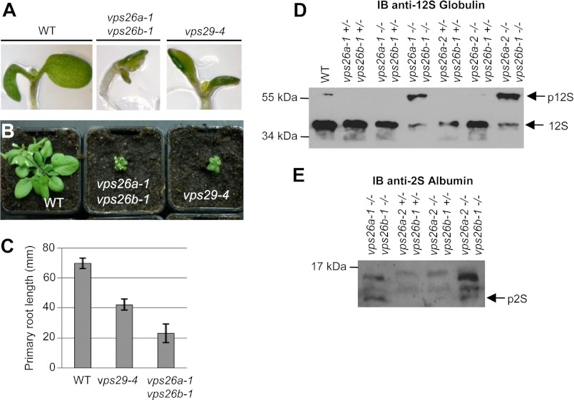
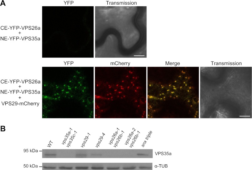
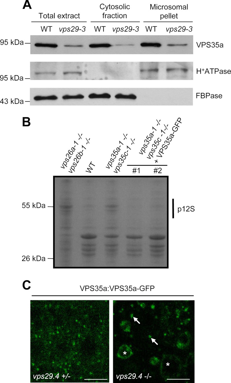
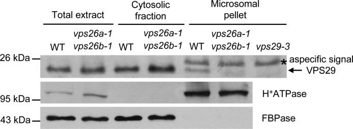
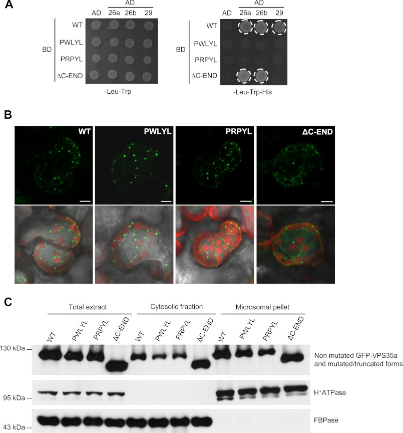
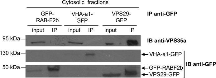
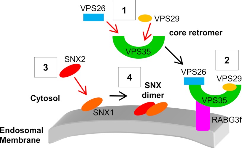
The retromer complex localizes to endosomal membranes and is involved in protein trafficking. In mammals, it is composed of a dimer of sorting nexins and of the core retromer consisting of vacuolar protein sorting (VPS)26, VPS29, and VPS35. Although homologs of these proteins have been identified in plants, how the plant retromer functions remains elusive. To better understand the role of VPS components in the assembly and function of the core retromer, we characterize here Arabidopsis vps26-null mutants. We show that impaired VPS26 function has a dramatic effect on VPS35 levels and causes severe phenotypic defects similar to those observed in vps29-null mutants. This implies that functions of plant VPS26, VPS29, and VPS35 are tightly linked. Then, by combining live-cell imaging with immunochemical and genetic approaches, we report that VPS35 alone is able to bind to endosomal membranes and plays an essential role in VPS26 and VPS29 membrane recruitment. We also show that the Arabidopsis Rab7 homolog RABG3f participates in the recruitment of the core retromer to the endosomal membrane by interacting with VPS35. Altogether our data provide original information on the molecular interactions that mediate assembly of the core retromer in plants.


Figures

FIGURE 1.
FIGURE 1.
vps26a vps26b double null mutants present defects similar to other vps mutants. A, cotyledon phenotype of 7-day-old seedlings of vps29-4 single and vps26a-1 vps26b-1 double mutants compared with wild type (WT). B, phenotype of adult WT, vps29-4 single, and vps26a-1 vps26b-1 double mutant plants. C, measures of primary root length of 5-day-old seedlings from WT, vps29-4 single, and vps26a-1 vps26b-1 double mutant plants. Differences are significant with a p value < 0.001. Error bars show S.E. D and E, total protein extracts from WT and various combinations of vps26a vps26b mutant seeds were analyzed by Western blotting using anti-12S globulin and anti-2S albumin antibodies. vps26a vps26b homozygous double mutants present abnormal accumulation of precursors of 12S-globulin (p12S) (D) and 2S-albumin (p2S) (E). 12S, mature 12S-globulin. Molecular mass markers are indicated on the left in kDa.
FIGURE 2.
FIGURE 2.
Stability of the core retromer. A, BiFC analysis of the interaction between NE-YFP-VPS35a and CE-YFP-VPS26a in epidermal cells of tobacco leaves in the absence (upper panel) or the presence of VPS29-mCherry (lower panel). In the absence of VPS29-mCherry, no interaction between VPS35a and VPS26a is detected. The YFP and mCherry channels, the superimposed image for the two channels (Merge), and images obtained with transmitted light (transmission) are shown. Scale bars, 10 μm. B, immunoblot analysis on total protein extracts from different Arabidopsis retromer mutants and wild-type (WT) plants using anti-VPS35a and anti-α-tubulin (α-TUB) antibodies. α-Tubulin was used as loading control. snx triple stands for snx1-2 snx2a-2 snx2b-1-null mutant. Molecular mass markers are indicated on the left in kDa.
FIGURE 3.
FIGURE 3.
VPS29 is dispensable for VPS35 membrane recruitment. A, immunoblot analysis on total, cytosolic, and microsomal proteins from wild-type (WT) and vps29-3 plantlets using anti-VPS35a, anti-H+-ATPase, and anti-cytosolic FBPase antibodies. H+-ATPase and FBPase were used as markers of membrane and cytosolic fractions, respectively. Molecular mass markers are indicated on the left in kDa. B, total proteins extracted from dry seeds of wild type (WT), vps26a vps26b, vps35a vps35c, and two transgenic vps35a vps35c independent lines (#1 and #2) stably expressing the VPS35a-GFP fusion protein under the pVPS35a promoter. Extracts were analyzed by SDS-PAGE followed by Coomassie Blue staining of the gel. Transgenic lines exhibit a WT-like pattern of seed storage proteins compared with the mutants. p12S: precursor of 12S-globulin. Molecular mass markers are indicated on the left in kDa. C, confocal microscopy images illustrating the localization of VPS35a-GFP in the roots of vps29-4+/− (left) and vps29–4−/− (right) plants. Arrows indicate punctate compartments, and asterisks point to abnormally enlarged/vacuolated compartments labeled with VPS35a-GFP. Scale bars, 10 μm.
FIGURE 4.
FIGURE 4.
VPS26 is required for VPS29 endosomal recruitment. Immunoblot analysis of total, cytosolic, and microsomal proteins from wild-type (WT) and vps26a-1 vps26b-1 plants using anti-VPS29, anti-H+-ATPase, and anti-cytosolic FBPase antibodies. H+-ATPase and FBPase were used as markers of membrane and cytosolic fractions, respectively. The arrow corresponds to VPS29, and the asterisk indicates a protein that is nonspecifically recognized by the anti-VPS29 antibody in microsomal fraction because it is detected in vps29-3 mutant (top panel). Molecular mass markers are indicated on the left in kDa.
FIGURE 5.
FIGURE 5.
VPS35 binds to membranes independently of VPS26 and VPS29. A, yeast two-hybrid test monitoring the interaction between VPS26a, VPS26b, VPS29, and the wild-type (WT) and mutated/truncated forms of VPS35a. The corresponding empty vectors were used as negative controls. The interaction is revealed by the activation of HIS3 transcription and growth on -His medium and is indicated by a white circle. The mutated VPS35a forms lose their capacity to bind to VPS26a, VPS26b, and VPS29, whereas the truncated VPS35a deleted of its C terminus can still associate with VPS26a and VPS26b, but not with VPS29. PWLYL, VPS35a-R104W; PRPYL, VPS35a-L105P; ΔC-END, VPS35a-ΔC-END; 26a, VPS26a; 26b, VPS26b; 29, VPS29; AD, Gal4 activation domain; BD, Gal4 binding domain. B, confocal microscopy analysis of the localization of WT, mutated (PWLYL, PRPYL), and C-terminal truncated (ΔC-END) forms of VPS35a tagged with GFP in N. benthamiana mesophyll leaf cells. Pictures below correspond to the overlay of GFP signals and images obtained with transmitted light. Chlorophyll autofluorescence in chloroplasts is seen in red. Scale bars, 10 μm. C, immunoblot analysis of total, cytosolic, and microsomal proteins extracted from tobacco leaves expressing GFP-tagged VPS35a or its mutated/truncated forms using anti-GFP, anti-H+-ATPase, and anti-cytosolic FBPase antibodies. H+-ATPase and FBPase were used as markers of membrane and cytosolic fractions, respectively. Molecular mass markers are indicated on the left in kDa. WT, wild-type VPS35a fused to GFP; PWLYL, GFP-VPS35a-R104W; PRPYL, GFP-VPS35a-L105P; ΔC-END, C-terminal truncated form of VPS35a fused to GFP.
FIGURE 6.
FIGURE 6.
VPS29 and VPS35 are able to associate in the cytosol. Co-IP of VPS29-GFP and VPS35a was performed with an anti-GFP antibody on soluble fractions of transgenic plantlets expressing VPS29-GFP. Transgenic plantlets expressing either GFP-RabF2b or VHA-a1-GFP were used as negative controls for the IP assay. VPS35a and GFP-tagged proteins were detected with anti-VPS35a and anti-GFP antibodies, respectively. IB, immunoblotting. Molecular mass markers are indicated on the left in kDa.
FIGURE 7.
FIGURE 7.
VPS35a associates with RABG3f on membranes. A, co-localization of mCherry-VPS35a with GFP-RABG3f in endosomal compartments. Confocal analysis was performed on epidermal cells of tobacco leaves co-expressing the two fusion proteins. Scale bars, 10 μm. B, yeast two-hybrid test monitoring the interaction between VPS35a and RABG3f. The corresponding empty vectors were used as negative controls. Yeast transformants were grown in liquid cultures and diluted to A600 nm = 0.2, 0.02, and 0.002 and then spotted on −Leu −Trp (left panel) and −Leu −Trp −His (right panel) synthetic complete (SC) medium. The interaction is revealed by the activation of HIS3 transcription and growth on −His medium. AD, Gal4 activation domain; BD, Gal4 DNA binding domain. C, co-IP of YFP-RABG3f and VPS35a with an anti-GFP antibody from microsomal protein extracts of transgenic plantlets expressing YFP-RABG3f. Transgenic plantlets expressing the partially membrane localized protein 2× mCitrine containing a myristoylation tail (Myr-2× m-Cit), as well as wild-type (WT) plantlets were used to test the specificity of the YFP-RABG3f/VPS35a interaction. VPS35a and GFP-tagged proteins were detected with anti-VPS35a and anti-GFP antibodies, respectively. The asterisk indicates a protein that is nonspecifically recognized by the anti-GFP antibody in microsomal fraction. M, microsomal pellet (Input); IP, immunoprecipitate; IB, immunoblotting. D, immunoprecipitation (IP) assays from cytosolic fractions of YFP-RABG3f and Myr-2× m-Cit transgenic and WT plants reveal that VPS35a does not interact with YFP-RABG3f in the soluble fraction. IP and immunodetection were conducted as in C. S, soluble fractions (Input).
FIGURE 8.
FIGURE 8.
Tentative model of the assembly of the core retromer to endosomal membranes in plants. The core retromer assembles first in the cytosol (1) and then is recruited to the endosomal membrane through an interaction between VPS35 and RABG3f (2). This recruitment occurs independently of SNX proteins. SNXs participate in certain functions of the core retromer through the formation of membrane-bound heterodimers consisting of SNX1 and SNX2s. In plants, SNX1 is the only SNX capable of binding to the membrane of endosomes (3) and will recruit cytosolic SNX2s to form a functional SNX complex (4).

Similar articles

Cited by

References

    1. Attar N., Cullen P. J. (2010) The retromer complex. Adv. Enzyme Regul. 50, 216–236 - PubMed
    1. Griffin C. T., Trejo J., Magnuson T. (2005) Genetic evidence for a mammalian retromer complex containing sorting nexins 1 and 2. Proc. Natl. Acad. Sci. U.S.A. 102, 15173–15177 - PMC - PubMed
    1. Wassmer T., Attar N., Bujny M. V., Oakley J., Traer C. J., Cullen P. J. (2007) A loss-of-function screen reveals SNX5 and SNX6 as potential components of the mammalian retromer. J. Cell Sci. 120, 45–54 - PubMed
    1. Wassmer T., Attar N., Harterink M., van Weering J. R., Traer C. J., Oakley J., Goud B., Stephens D. J., Verkade P., Korswagen H. C., Cullen P. J. (2009) The retromer coat complex coordinates endosomal sorting and dynein-mediated transport, with carrier recognition by the trans-Golgi network. Dev. Cell 17, 110–122 - PMC - PubMed
    1. Nothwehr S. F., Bruinsma P., Strawn L. A. (1999) Distinct domains within Vps35p mediate the retrieval of two different cargo proteins from the yeast prevacuolar/endosomal compartment. Mol. Biol. Cell 10, 875–890 - PMC - PubMed

Publication types

MeSH terms

Substances