. 2013 Mar 22;288(12):8585-8595.
doi: 10.1074/jbc.M112.413997. Epub 2013 Jan 28.

Dentin phosphophoryn activates Smad protein signaling through Ca2+-calmodulin-dependent protein kinase II in undifferentiated mesenchymal cells

Affiliations

Dentin phosphophoryn activates Smad protein signaling through Ca2+-calmodulin-dependent protein kinase II in undifferentiated mesenchymal cells

Asha Eapen et al. J Biol Chem. .

Abstract

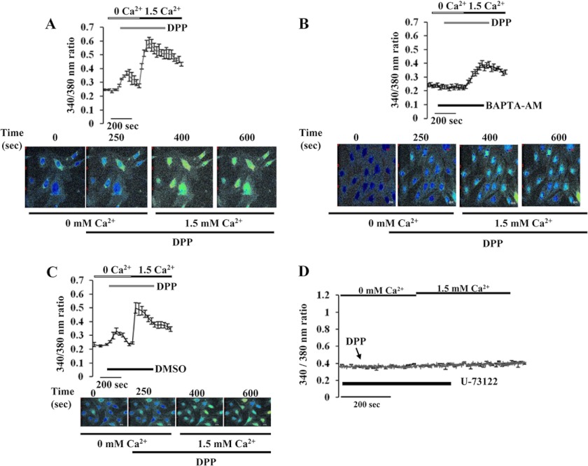
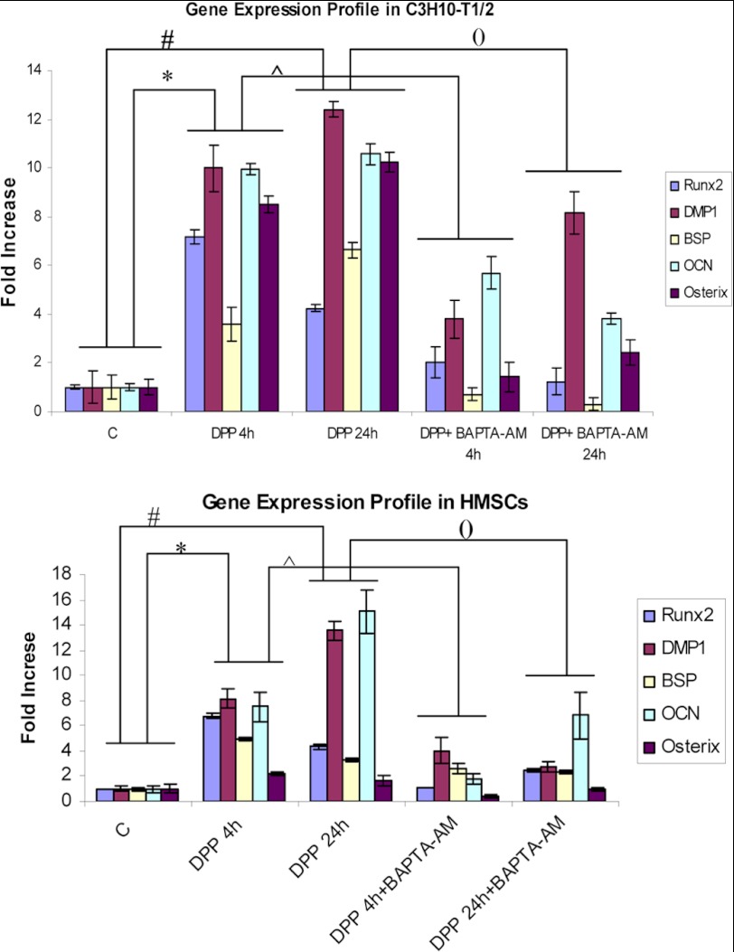
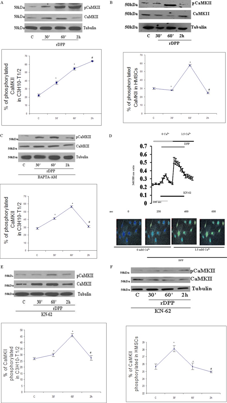
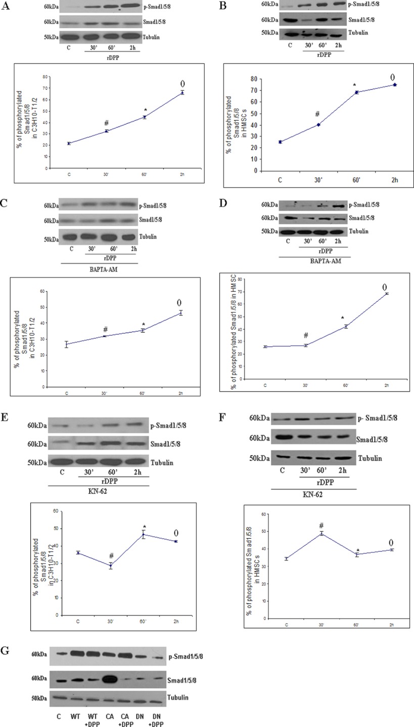
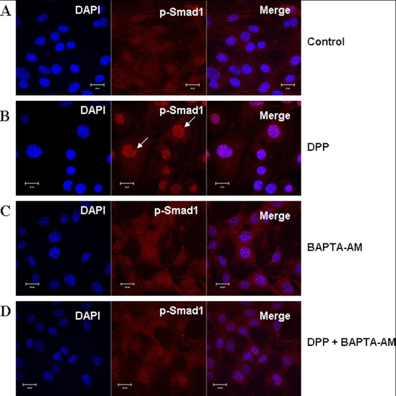
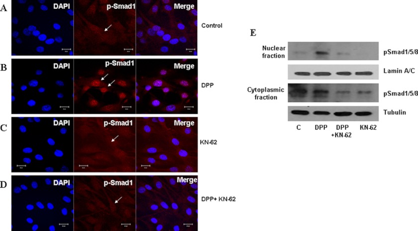
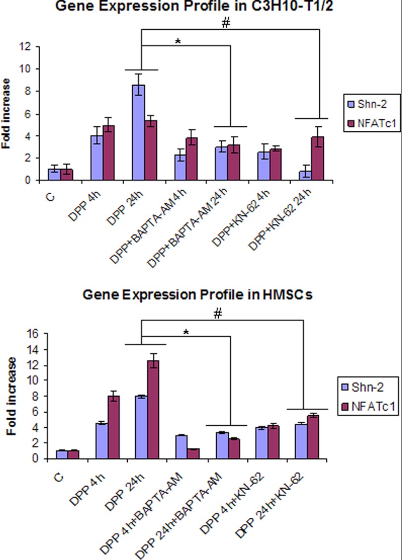
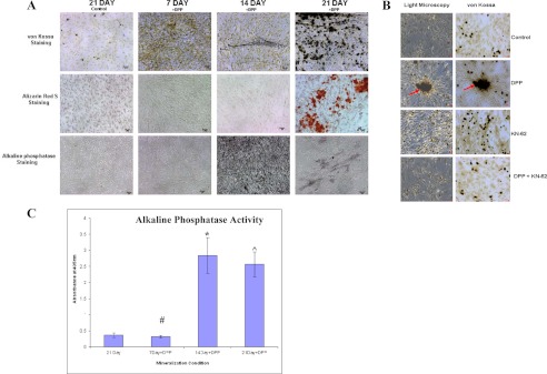
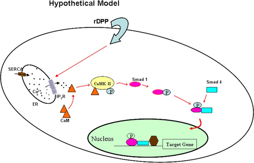
Dentin phosphophoryn (DPP) is a major noncollagenous protein in the dentin matrix. In this study, we demonstrate that pluripotent stem cells such as C3H10T1/2 and human bone marrow cells can be committed to the osteogenic lineage by DPP. Treatment with DPP can stimulate the release of intracellular Ca(2+). This calcium flux triggered the activation of Ca(2+)-calmodulin-dependent protein kinase II (CaMKII). Activated CaMKII induced the phosphorylation of Smad1 and promoted nuclear translocation of p-Smad1. Inhibition of store Ca(2+) depletion by 1,2-bis(2-aminophenoxy)ethane-N,N,N',N'-tetraacetic acid tetrakis(acetoxymethyl ester) or down-regulation of CaMKII by KN-62, a selective cell-permeable pharmacological inhibitor or a dominant negative plasmid of CaMKII, blocked DPP-mediated Smad1 phosphorylation. Activation of Smad1 resulted in the expression of osteogenic markers such as Runx2, Osterix, DMP1, Bone sialoprotein, Osteocalcin, NFATc1, and Schnurri-2, which have been implicated in osteoblast differentiation. These findings suggest that DPP is capable of triggering commitment of pluripotent stem cells to the osteogenic lineage.


Figures

FIGURE 1.
FIGURE 1.
A, effect of rDPP on [Ca2+]i in mesenchymal stem cells. DPP-induced [Ca2+]i was measured in C3H10T1/2 cells as described under “Materials and Methods.” Fura-2AM-loaded cells were washed three times, placed in Ca2+-free and Mg2+-free HBSS, and then stimulated with rDPP (500 ng/ml). Experiments were repeated three times, and a representative plot is shown. Picture shots display DPP-induced [Ca2+]i release in C3H10T1/2 cells at 250 s. Yellow color within the cells indicates the release of calcium from the intracellular stores when C3H10T1/2 cells were stimulated with DPP or the intake of calcium upon depletion of the stores when extracellular calcium was added. B, BAPTA-AM is a calcium chelator and can block DPP-stimulated increase in [Ca2+]i in C3H10T1/2 cells. Cells pretreated with BAPTA-AM (50 μm) were loaded with Fura-2AM and placed in Ca2+- and Mg2+-free HBSS. The cells were then stimulated with rDPP (500 ng/ml) and then observed for Ca2+ store release. Cells with no BAPTA-AM treatment served as control. Experiments were performed in triplicate. A representative plot is shown in this panel. Picture shots display DPP-induced release in BAPTA-AM-pretreated C3H10T1/2 cells at 250 s. C, DMSO treatment does not inhibit DPP-induced [Ca2+]i release in C3H10T1/2 cells. Cells pretreated with DMSO (0.01%) were loaded with Fura-2AM and placed in Ca2+- and Mg2+-free HBSS. The cells were then stimulated with rDPP (500 ng/ml) and then observed for Ca2+ store release. As DMSO was used as a solvent vehicle to dissolve all inhibitors, it was therefore necessary to identify if the vehicle had an effect on [Ca2+]i release in C3H10T1/2 cells. Screen shots of the live image of DPP induced [Ca2+]i release in DMSO-pretreated C3H10T1/2 cells. D, U-73122 inhibitor of the phospholipase C pathway inhibits DPP-induced [Ca2+]i release in C3H10T1/2 cells. DPP-induced [Ca2+]i was measured in confluent mesenchymal stem cells upon stimulation with 500 ng/ml rDPP after 30 min of pretreatment with U-73122 (phospholipase C inhibitor). Cells were loaded with Fura-2AM and placed in Ca2+- and Mg2+-free HBSS and stimulated with rDPP (500 ng/ml) to measure store-Ca2+ release. Arrow indicates the time at which cells were stimulated with rDPP. Experiments were repeated a minimum of four times. A representative plot is shown in this panel.
FIGURE 2.
FIGURE 2.
DPP enhances osteoblastic gene expression in mesenchymal stem cells. C3H10T1/2 cells and HMSCs in basal media for 24 h were stimulated with 500 ng/ml rDPP with or without BAPTA-AM treatment for 4 and 24 h. Total RNA was isolated and subjected to real time PCR and analyzed for gene expression of Runx2, BSP, DMP1, Osterix, and OCN. Untreated C3H10T1/2 cells served as control. These results were normalized with the loading control GAPDH. Experiments were done in triplicate. *, #, (), and [caret], p < 0.05. C, control.
FIGURE 3.
FIGURE 3.
A, DPP activates CaMKII in C3H10T1/2 cells. C3H10T1/2 cells were stimulated with rDPP (500 ng/ml), and Western blotting was performed. Experiments were done in triplicate. *, #, and [caret], p < 0.05 as compared with untreated cells. B, DPP activates CaMKII in HMSCs. C3H10T1/2 cells were stimulated with rDPP (500 ng/ml), and Western blotting was performed with Abs against CaMKII. Experiments were done in triplicate. *, #, and [caret], p < 0.05 as compared with untreated cells. C, DPP-mediated activation of CaMKII can be inhibited by the treatment with BAPTA-AM. C3H10T1/2 cells were pretreated with BAPTA-AM (50 μm) followed by stimulation with rDPP for the indicated time points, and Western blotting was performed with Abs against CaMKII. Experiments were done in triplicate. *, #, and [caret], p < 0.05 as compared with untreated cells. D, CaMKII inhibitor KN-62 inhibits DPP-induced [Ca2+]i release in C3H10T1/2 cells. DPP-induced [Ca2+]i was measured in confluent C3H10T1/2 cells loaded with 10 μm KN-62 for 30 min prior to the addition of Fura-2AM. Fura-2AM-loaded cells were washed three times, placed in Ca2+- and Mg2+-free HBSS, and then stimulated with rDPP (500 ng/ml). The experiment was repeated four times, and a representative plot is shown. Picture images display DPP-induced [Ca2+]i release in KN-62-pretreated C3H10T1/2 cells at 250 s. E, KN-62 inhibits CaMKII activation in C3H10T1/2 cells. Treatment of C3H10T1/2 cells with KN-62 (10 μm) inhibits CaMKII activation. Total proteins were isolated, and Western blots were developed with CaMKII antibody and anti-phospho-CaMKII antibody. Equal protein loading was confirmed by stripping the blot followed by probing it with tubulin antibody. Experiments were done in triplicate. *, #, and [caret], p < 0.05 as compared with untreated cells. F, KN-62 inhibits DPP-mediated phosphorylation of CaMKII in HMSCs. Total proteins were isolated from DPP-stimulated HMSCs, and immunoblots were performed with CaMKII antibody and anti-phospho-CaMKII antibody. Experiments were done in triplicate. *, #, and [caret], p < 0.05 as compared with untreated cells. C, control.
FIGURE 4.
FIGURE 4.
Activation of Smad1/5/8 in C3H10T1/2 and HMSCs by DPP can be inhibited by the treatment with BAPTA-AM and KN-62. A, C3H10T1/2 cells were stimulated with rDPP (500 ng/ml), and Western blotting was performed with Abs against Smad1/5/8. *, #, and (), p < 0.05 as compared with control cells. B, HMSCs were stimulated with rDPP (500 ng/ml), and Western blotting was performed with Abs against Smad1/5/8. *, #, and (), p < 0.05 as compared with control cells. C, C3H10T1/2 cells were pretreated with BAPTA-AM (50 μm) followed by stimulation with rDPP for the indicated time points, and Western blotting was performed with Abs against Smad1/5/8. *, #, and (), p < 0.05 as compared with control cells. D, HMSCs were treated with BAPTA-AM (50 μm) followed by stimulation with rDPP, and Western blotting was performed with Abs against Smad1/5/8. *, #, and (), p < 0.05 as compared with control cells. E, treatment of C3H10T1/2 cells with KN-62 (10 μm) inhibits Smad1/5/8 activation. Total proteins were isolated, and Western blots were developed with anti-Smad1/5/8 antibody and anti-phospho-Smad1/5/8 antibody. Equal loading of the proteins was confirmed by stripping the blot followed by probing it with tubulin. *, #, and (), p < 0.05 as compared with control cells. F, inhibition of Smad1/5/8 in KN-62-treated HMSCs, and Western blotting was performed with Abs against Smad1/5/8. *, #, and (), p < 0.05 as compared with control cells. G, C3H10T1/2 cells were transduced with wild type (WT), constitutively active (CA), and dominant negative (DN) CaMKII and then stimulated with rDPP. Western blotting was performed with Abs against Smad1/5/8. C, control.
FIGURE 5.
FIGURE 5.
Nuclear localization of p-Smad1 in C3H10T1/2 cells stimulated by DPP and abrogated by BAPTA-AM. C3H10T1/2 cells were stimulated with rDPP (500 ng/ml) or BAPTA-AM (50 μm) for 1 h. The cells were fixed and immunostained for p-Smad1 (red). Control cells exhibited diffused staining throughout the cells for p-Smad1 (A). Upon stimulation with DPP for 1 h, nuclear translocation of p-Smad1 was observed. Arrows indicate nuclear localization of p-Smad1 (B). Cells treated with BAPTA-AM alone displayed cytoplasmic staining of p-Smad1 (C). However, DPP-stimulated and BAPTA-AM-pretreated cells failed to show nuclear translocation of p-Smad1 (D). DAPI (blue) was used to stain the nucleus. Bar, 10 μm.
FIGURE 6.
FIGURE 6.
DPP-mediated nuclear localization of p-Smad1 in C3H10T1/2 is abrogated in the presence of KN-62. C3H10T1/2 cells were stimulated with rDPP (500 ng/ml) for 1 h. Cells were fixed and immunostained for p-Smad1. Immunofluorescence shows p-Smad1 predominantly located in the cytoplasm of untreated cells (A). With DPP stimulation, the predominant nuclear localization of p-Smad1 is observed. Arrows indicate nuclear localization of p-Smad1 (B). Diffused cytoplasmic staining of p-Smad1 was observed in cells pretreated with KN-62 alone (C). However, C3H10T1/2 cells stimulated with rDPP and KN-62 prevented nuclear localization of p-Smad1 (D). Bar, 10 μm. Nuclear and cytoplasmic proteins were extracted from C3H10T1/2 cells stimulated with DPP, and Western blotting was performed with anti-p-Smad1/5/8 and Smad1/5/8 antibody. The blots were stripped and reprobed with lamin A/C and tubulin (E). C, control.
FIGURE 7.
FIGURE 7.
DPP activates Smad pathway-related gene expression. C3H10T1/2 and HMSCs were stimulated with rDPP (500 ng/ml) or KN-62 (10 μm) or BAPTA-AM (50 μm) for 4 and 24 h, respectively. Total RNA was extracted, and expression levels of NFATc1 and SHN2 were estimated based on real time PCR. Upon stimulation with DPP, expression levels of these genes increased from 4 to 24 h. Lower expression levels were observed with KN-62 and BAPTA-AM pretreatment. Experiments were performed in triplicate. * and #, p < 0.05. C, control.
FIGURE 8.
FIGURE 8.
DPP stimulates mineralized nodule formation. C3H10T1/2 cells were cultured in mineralization media for 30 days either with rDPP (500 ng/ml) or KN-62 (10 μm) or both. von Kossa, Alizarin Red S, and alkaline phosphatase staining were performed (A). Terminal differentiation and mineralized nodule formation were observed when cells were stimulated with rDPP. However, untreated control cells, cells treated with inhibitor alone, and cells treated with the inhibitor in the presence of DPP did not show evidence of mineralized nodule formation (B). Arrows point to the mineralized nodule formed. Alkaline phosphatase activity was highly expressed at 14 and 21 days in DPP-treated cells (C). *, #, and [caret], p < 0.05 as compared with control cells. Bar, 20 μm.
FIGURE 9.
FIGURE 9.
Hypothetical model. Model showing DPP-mediated intracellular calcium store flux can trigger the activation of CaMKII, resulting in the activation of Smad1/5/8 signaling cascade. ER, endoplasmic reticulum; SERCA, sarco/endoplasmic reticulum Ca2+-ATPase; IP3R, inositol 1,4,5-trisphosphate receptor.

Similar articles

Cited by

References

    1. Veis A., Perry A. (1967) The phosphoprotein of the dentin matrix. Biochemistry 6, 2409–2416 - PubMed
    1. Ogbureke K. U., Fisher L. W. (2004) Expression of SIBLINGs and their partner MMPs in salivary glands. J. Dent. Res. 83, 664–670 - PubMed
    1. Linde A., Bhown M., Butler W. T. (1980) Noncollagenous proteins of dentin. A re-examination of proteins from rat incisor dentin utilizing techniques to avoid artifacts. J. Biol. Chem. 255, 5931–5942 - PubMed
    1. Richardson W. S., Munksgaard E. C., Butler W. T. (1978) Rat incisor phosphoprotein. J. Biol. Chem. 253, 8042–8046 - PubMed
    1. Feng J. Q., Luan X., Wallace J., Jing D., Ohshima T., Kulkarni A. B., D'Souza R. N., Kozak C. A., MacDougall M. (1998) Genomic organization, chromosomal mapping, and promoter analysis of the mouse dentin sialophosphoprotein (Dspp) gene, which codes for both dentin sialoprotein and dentin phosphoprotein. J. Biol. Chem. 273, 9457–9464 - PubMed

Publication types

MeSH terms

LinkOut - more resources