Islet β cell mass in diabetes and how it relates to function, birth, and death
- PMID: 23363033
- PMCID: PMC3618572
- DOI: 10.1111/nyas.12031
Islet β cell mass in diabetes and how it relates to function, birth, and death
Abstract
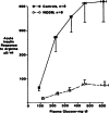
In type 1 diabetes (T1D) β cell mass is markedly reduced by autoimmunity. Type 2 diabetes (T2D) results from inadequate β cell mass and function that can no longer compensate for insulin resistance. The reduction of β cell mass in T2D may result from increased cell death and/or inadequate birth through replication and neogenesis. Reduction in mass allows glucose levels to rise, which places β cells in an unfamiliar hyperglycemic environment, leading to marked changes in their phenotype and a dramatic loss of glucose-stimulated insulin secretion (GSIS), which worsens as glucose levels climb. Toxic effects of glucose on β cells (glucotoxicity) appear to be the culprit. This dysfunctional insulin secretion can be reversed when glucose levels are lowered by treatment, a finding with therapeutic significance. Restoration of β cell mass in both types of diabetes could be accomplished by either β cell regeneration or transplantation. Learning more about the relationships between β cell mass, turnover, and function and finding ways to restore β cell mass are among the most urgent priorities for diabetes research.
© 2013 New York Academy of Sciences.
Figures


References
-
- Maclean N, Ogilvie RF. Quantitative estimation of the pancreatic islet tissue in diabetic subjects. Diabetes. 1955;4:367–376. - PubMed
-
- Butler AE, et al. Beta-cell deficit and increased beta-cell apoptosis in humans with type 2 diabetes. Diabetes. 2003;52:102–110. - PubMed
-
- Yoon KH, et al. Selective beta-cell loss and alpha-cell expansion in patients with type 2 diabetes mellitus in Korea. J. Clin. Endocrinol. Metab. 2003;88:2300–2308. - PubMed
-
- Rahier J, et al. Pancreatic beta-cell mass in European subjects with type 2 diabetes. Diabetes Obes. Metab. 2008;10(Suppl 4):32–42. - PubMed
-
- Kloppel G, et al. Islet pathology and the pathogenesis of type 1 and type 2 diabetes mellitus revisited. Surv. Synth. Pathol. Res. 1985;4:110–125. - PubMed
Publication types
MeSH terms
Grants and funding
LinkOut - more resources
Full Text Sources
Other Literature Sources
Medical

