. 2013 Jan 30:14:3.
doi: 10.1186/1471-2199-14-3.

UtroUp is a novel six zinc finger artificial transcription factor that recognises 18 base pairs of the utrophin promoter and efficiently drives utrophin upregulation

Affiliations

UtroUp is a novel six zinc finger artificial transcription factor that recognises 18 base pairs of the utrophin promoter and efficiently drives utrophin upregulation

Annalisa Onori et al. BMC Mol Biol. .

Abstract

Background: Duchenne muscular dystrophy (DMD) is the most common X-linked muscle degenerative disease and it is due to the absence of the cytoskeletal protein dystrophin. Currently there is no effective treatment for DMD. Among the different strategies for achieving a functional recovery of the dystrophic muscle, the upregulation of the dystrophin-related gene utrophin is becoming more and more feasible.

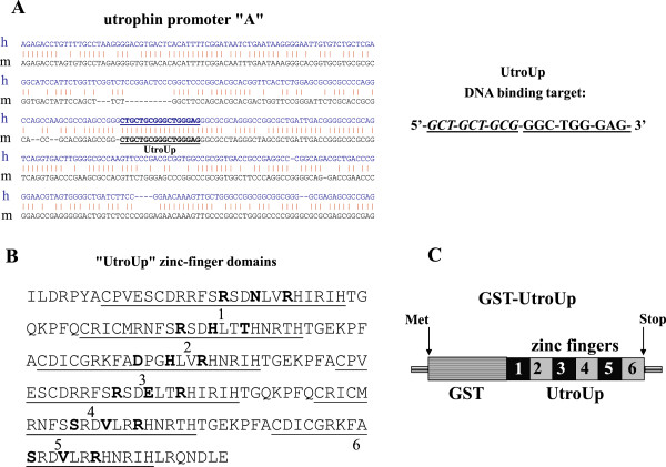
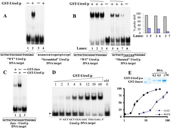
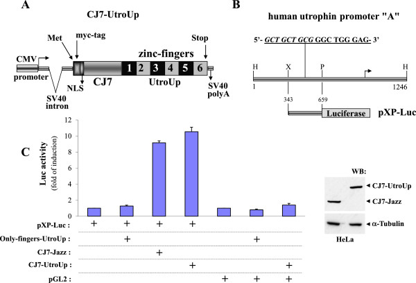
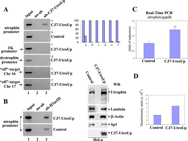
Results: We have previously shown that the zinc finger-based artificial transcriptional factor "Jazz" corrects the dystrophic pathology in mdx mice by upregulating utrophin gene expression. Here we describe a novel artificial transcription factor, named "UtroUp", engineered to further improve the DNA-binding specificity. UtroUp has been designed to recognise an extended DNA target sequence on both the human and mouse utrophin gene promoters. The UtroUp DNA-binding domain contains six zinc finger motifs in tandem, which is able to recognise an 18-base-pair DNA target sequence that statistically is present only once in the human genome. To achieve a higher transcriptional activation, we coupled the UtroUp DNA-binding domain with the innovative transcriptional activation domain, which was derived from the multivalent adaptor protein Che-1/AATF. We show that the artificial transcription factor UtroUp, due to its six zinc finger tandem motif, possesses a low dissociation constant that is consistent with a strong affinity/specificity toward its DNA-binding site. When expressed in mammalian cell lines, UtroUp promotes utrophin transcription and efficiently accesses active chromatin promoting accumulation of the acetylated form of histone H3 in the utrophin promoter locus.

Conclusions: This novel artificial molecule may represent an improved platform for the development of future applications in DMD treatment.


Figures

Figure 1
Figure 1
A graphical representation of the human and mouse utrophin promoters and the amino acid sequences of the six zinc finger UtroUp peptide (GenBank JQ073900). A: On the left is shown an alignment of DNA sequences of the human and mouse utrophin promoter “A”. The UtroUp DNA target sequence, which is 18 base pairs long, is underlined. The DNA target sequence is 100% conserved between humans and mice. On the right is shown the 18-base-pair UtroUp DNA target sequence. The portion of the Jazz DNA target sequence is indicated in italics. B: The amino acid sequence of the synthetic six zinc finger DNA-binding domain of the UtroUp peptide. Each zinc finger domain in the tandem array is underlined and numbered. The amino acid residues in the α-helix (−1, +3 and +6), which are key for the DNA-binding specificity, are indicated in bold. The zinc finger domains 4, 5 and 6 are inherited by the Jazz peptide. C: A schematic representation of the bacterially produced UtroUp peptide fused to glutathione S-transferase (GST).
Figure 2
Figure 2
The UtroUp DNA-binding affinity/specificity analysed by EMSA. A: An equal amount of the GST-UtroUp protein (14 nM) was incubated with an equal amount of either the labelled wild-type DNA target (lanes 1–2) or with the labelled scrambled DNA target (lanes 3–4). B: (Left) An increasing amount of the GST-UtroUp protein (20, 40, 80 nM) was incubated with an equal amount of either the labelled wild-type DNA target (lanes 1–4) or with the labelled mutant DNA target (lanes 5–8). The two mutated triplets in the DNA target are indicated by lowercase characters. (Right) Histogram shows a percentage of probes (WT and Mutated) shifted with increasing amounts of GST-UtroUp protein. C: An equal amount (12 nM), of the GST-UtroUp protein (lane 2) and GST-Jazz protein (lane 1) were incubated with an equal amount of labelled UtroUp-Jazz DNA target. D: An EMSA prototype performed for the determination of the dissociation constant (Kd). An increasing amount of GST-UtroUp protein (1, 2, 3, 4, 8, 12, 30, and 60 nM) was complexed with the labelled UtroUp DNA target. E: (Top) Quality control by Coomassie blue staining of purified/eluted GST-UtroUp and GST-Jazz fusion proteins compared with purified commercial bovine serum albumin. (Bottom) The Kd values of the GST-UtroUp/DNA-target and GST-Jazz/DNA complexes correspond approximately to 3,5 nM and 32 nM respectively. The protein concentration is expressed in nM.
Figure 3
Figure 3
The trans-activation of the human utrophin promoter “A”. A: Schematic representation of the CJ7-UtroUp chimera in the mammalian expression vector. B: A graphical representation of the human utrophin promoter “A”. The pXP-Luc construct consists of a minimal region of the human utrophin promoter “A” containing the 18-base-pair UtroUp target DNA-binding sequence (indicated in italicised and underlined characters) that was cloned upstream of the reporter gene (Luciferase). C: (Left) A histogram shows the fold of induction of the pXP-Luc utrophin promoter “A” construct or the control reporter pGL2-Promoter (Promega) that was obtained upon co-transfection with the finger region-only UtroUp, or the CJ7-UtroUp or the CJ7-Jazz in HeLa cells. The data are presented as the means ± S.D. of three independent experiments that were performed in triplicate. (Rigth) Western blot analysis cell lysates from HeLa cells transiently cotransfected with pXP-Luc construct and the indicated CJ7-UtroUp or CJ7-Jazz expression vectors. The membrane was incubated with the anti-myc tag antibody and the anti-alpha tubulin antibody.
Figure 4
Figure 4
The binding and upregulation of the endogenous utrophin gene. A: (Left) The ChIP assay was performed in transfected HeLa cells (with either an empty control vector or with the CJ7-UtroUp expression vector) using myc-tag monoclonal antibody/protein G-agarose beads. The immunoprecipitates from each sample were analysed by PCR performed using primers specific for the human utrophin promoter “A”. PCRs of the promoters of the unrelated thymidine kinase and dystrophin genes and PCRs of specific regions on the chromosomes 16 and 17 were included as controls. A sample representing the linear amplification of the total input chromatin (input) was included (lane 1). (Right) Histogram shows quantification of ChIP-PCR bands presented as fraction of the input. B: ChIP was performed in transfected HeLa cells (with either the CJ7-UtroUp expression vector or with an empty control vector) using anti-acetylated histone H3 rabbit polyclonal antibody/protein G-agarose beads. Immunoprecipitates from each sample were analyzed by PCR performed using primers specific for human utrophin promoter “A”. C: The analysis by real-time PCR of the utrophin gene mRNA expression in HeLa cells transfected with the CJ7-UtroUp or control vectors. The ratio between the utrophin and GAPDH gene expression is shown as means ± S.D. from three independent experiments that were performed in triplicate. D: (Left) Western blot analysis of total protein lysates from HeLa cells transfected with either empty vector (Control) or with the CJ7-UtroUp expression vector. The membrane was incubated with the indicated antibodies. (Right) Histogram shows utrophin expression evaluated by densitometric analysis.

Similar articles

Cited by

References

    1. McNally EM, Pytel P. Muscle Disease: The Muscular Dystrophies. Annu Rev Pathol Mech Dis. 2007;2:87–109. doi: 10.1146/annurev.pathol.2.010506.091936. - DOI - PubMed
    1. Fairclough RJ, Bareja A, Davies KE. Progress in therapy for Duchenne muscular dystrophy. Exp Physiol. 2011;96(11):1101–1113. doi: 10.1113/expphysiol.2010.053025. - DOI - PubMed
    1. Pichavant C, Aartsma-Rus A, Clemens PR, Davies KE, Dickson G. et al.Current status of pharmaceutical and genetic therapeutic approaches to treat DMD. Mol Ther. 2011;19(5):830–840. doi: 10.1038/mt.2011.59. - DOI - PMC - PubMed
    1. Goyenvalle A, Seto JT, Davies KE, Chamberlain J. Therapeutic approaches to muscular dystrophy. Hum Mol Genet. 2011;20(R1):R69–78. doi: 10.1093/hmg/ddr105. - DOI - PMC - PubMed
    1. Meng J, Muntoni F, Morgan JE. Stem cells to treat muscular dystrophies - where are we? Neuromuscul Disord. 2011;21(1):4–12. doi: 10.1016/j.nmd.2010.10.004. - DOI - PubMed

Publication types