. 2013 Jan 30:11:2.
doi: 10.1186/1477-3155-11-2.

3, 4-dihydroxy-L-phenylalanine-derived melanin from Yarrowia lipolytica mediates the synthesis of silver and gold nanostructures

Affiliations

3, 4-dihydroxy-L-phenylalanine-derived melanin from Yarrowia lipolytica mediates the synthesis of silver and gold nanostructures

Mugdha Apte et al. J Nanobiotechnology. .

Abstract

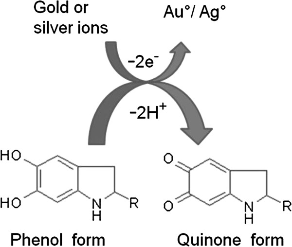
Background: Nanobiotechnology applies the capabilities of biological systems in generating a variety of nano-sized structures. Plants, algae, fungi and bacteria are some systems mediating such reactions. In fungi, the synthesis of melanin is an important strategy for cell-survival under metal-stressed conditions. Yarrowia lipolytica, the biotechnologically significant yeast also produces melanin that sequesters heavy metal ions. The content of this cell-associated melanin is often low and precursors such as L-tyrosine or 3, 4-dihydroxy-L-phenylalanine (L-DOPA) can enhance its production. The induced melanin has not been exploited for the synthesis of nanostructures. In this investigation, we have employed L-DOPA-melanin for the facile synthesis of silver and gold nanostructures. The former have been used for the development of anti-fungal paints.

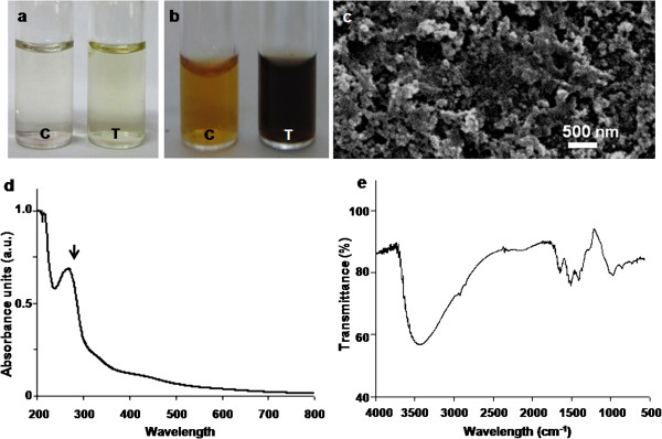
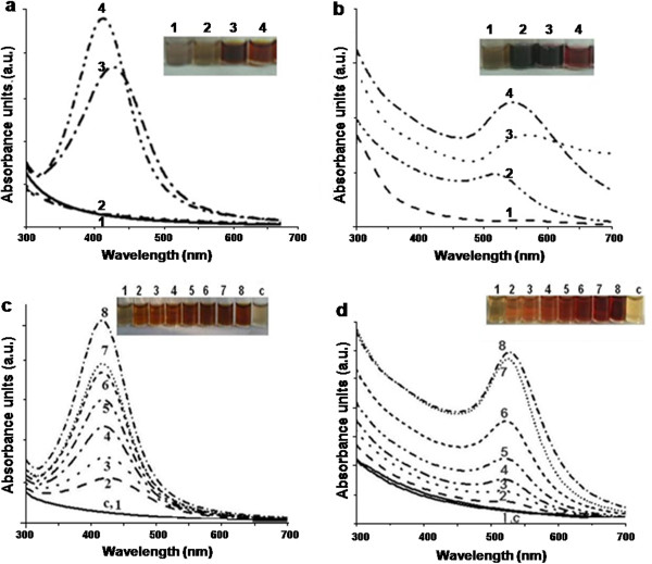
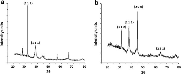
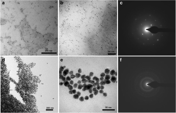
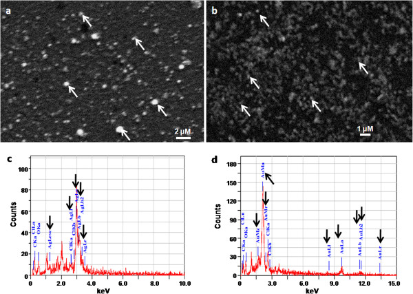
Methods: Yarrowia lipolytica NCIM 3590 cells were incubated with L-DOPA for 18 h and the resultant dark pigment was subjected to physical and chemical analysis. This biopolymer was used as a reducing and stabilizing agent for the synthesis of silver and gold nanostructures. These nanoparticles were characterized by UV-Visible spectra, X-ray diffraction (XRD) studies, and electron microscopy. Silver nanoparticles were evaluated for anti-fungal activity.

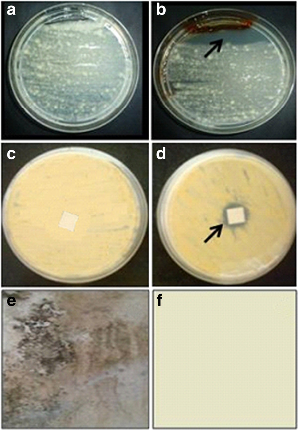
Results: The pigment isolated from Y. lipolytica was identified as melanin. The induced pigment reduced silver nitrate and chloroauric acid to silver and gold nanostructures, respectively. The silver nanoparticles were smaller in size (7 nm) and displayed excellent anti-fungal properties towards an Aspergillus sp. isolated from a wall surface. An application of these nanoparticles as effective paint-additives has been demonstrated.

Conclusion: The yeast mediated enhanced production of the metal-ion-reducing pigment, melanin. A simple and rapid method for the extracellular synthesis of nanoparticles with paint-additive-application was developed.


Figures

Figure 1
Figure 1
Characterization of the L-DOPA induced-melanin derived from Y. lipolytica NCIM 3590. Visual observations of reaction mixtures containing (a) L-tyrosine (b) L-DOPA [in Figures a and b, tube C depicts control tube without cells and tube T depicts reaction mixtures with resting cells]. Representative (c) SEM image (d) UV-Visible spectrum (e) FTIR spectrum.
Figure 2
Figure 2
Effect of pH on the synthesis of (a) silver and (b) gold nanoparticles by L-DOPA induced-melanin derived from Y. lipolytica NCIM 3590. [Reaction mixtures (2 ml) contained 10 μg of melanin at different pH, 1 mM metal salt incubated at 100°C for 10 min. Tubes and lines 1–4 depict pH of 6.0, 8.0, 10.0 and 12.0, respectively]. Effect of salt concentration on the synthesis of (c) silver and (d) gold nanoparticles [Reaction mixtures (2 ml) contained 10 μg of melanin at pH 12.0 and different concentrations of metal salts incubated at 100°C for 10 min. Tubes and lines 1–8 depict salt concentrations of 0.25, 0.5, 0.75, 1.0, 1.25, 1.5, 1.75 and 2.0 mM, respectively; tube and line C depicts a control without melanin].
Figure 3
Figure 3
Representative XRD profiles of melanin-mediated nanoparticles of (a) silver (b) gold. Three peaks in Figure (a) are unassigned.
Figure 4
Figure 4
Representative TEM images (at different magnifications) and SAED profiles of melanin-mediated nanoparticles of (a, b and c) silver and (d, e and f) gold, respectively.
Figure 5
Figure 5
Representative SEM images of melanin-induced (a) silver and (b) gold nanoparticles. Representative EDS profiles of (c) silver and (d) gold nanoparticles.
Figure 6
Figure 6
Proposed mechanism involved in the synthesis of nanoparticles by L-DOPA induced-melanin derived from Y. lipolytica.
Figure 7
Figure 7
Anti-fungal activity of silver nanoparticles synthesized by the L-DOPA induced-melanin derived from Y. lipolytica. Plates depicting growth of Aspergillus sp. (a) without (b) with silver nanoparticles. Paint-coated filter paper strips (c) without and (d) with silver nanoparticles as seen from the base of the plate. Representative pictures depicting the growth of the fungus on glass plates coated with (e) un-modified and (f) modified paint.

References

    1. Li X, Xu H, Chen ZS, Chen G. Biosynthesis of nanoparticles by microorganisms and their applications. J Nanomater. 2011. - DOI
    1. Mandal D, Bolander ME, Mukhopadhyay D, Sarkar G, Mukherjee P. The use of microorganisms for the formation of metal nanoparticles and their application. Appl Microbiol Biotechnol. 2006;69:485–492. doi: 10.1007/s00253-005-0179-3. - DOI - PubMed
    1. Duran N, Marcato PD, Duran M, Yadav A, Gade A, Rai M. Mechanistic aspects in the biogenic synthesis of extracellular metal nanoparticles by peptides, bacteria, fungi, and plants. Appl Microbiol Biotechnol. 2011;90:1609–1624. doi: 10.1007/s00253-011-3249-8. - DOI - PubMed
    1. Mohanpuria P, Rana N, Yadav SK. Biosynthesis of nanoparticles: technological concepts and future applications. J Nanopart Res. 2008;10:507–517. doi: 10.1007/s11051-007-9275-x. - DOI
    1. Thakkar KN, Mhatre SS, Parikh RY. Biological synthesis of metallic nanoparticles. Nanomed: Nanotechnol Biol Med. 2010;6:257–262. doi: 10.1016/j.nano.2009.07.002. - DOI - PubMed

Publication types

MeSH terms