. 2013 Apr;24(7):923-32.
doi: 10.1091/mbc.E13-01-0034. Epub 2013 Jan 30.

Assembly in G1 phase and long-term stability are unique intrinsic features of CENP-A nucleosomes

Affiliations

Assembly in G1 phase and long-term stability are unique intrinsic features of CENP-A nucleosomes

Dani L Bodor et al. Mol Biol Cell. 2013 Apr.

Abstract

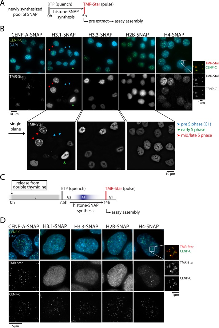
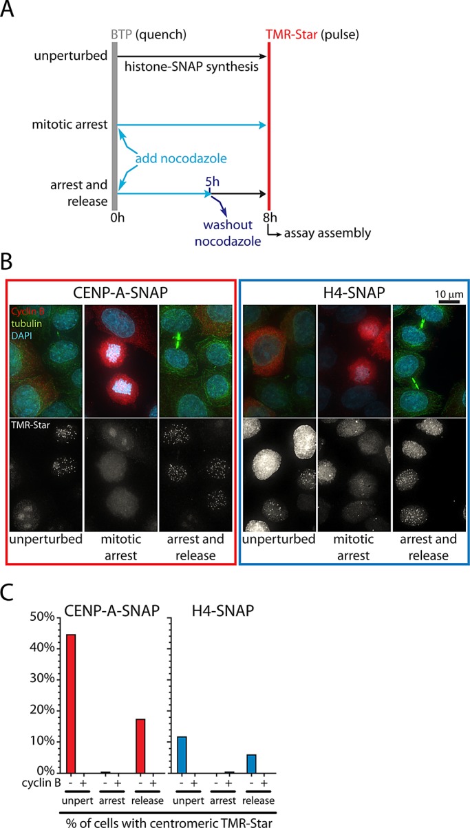
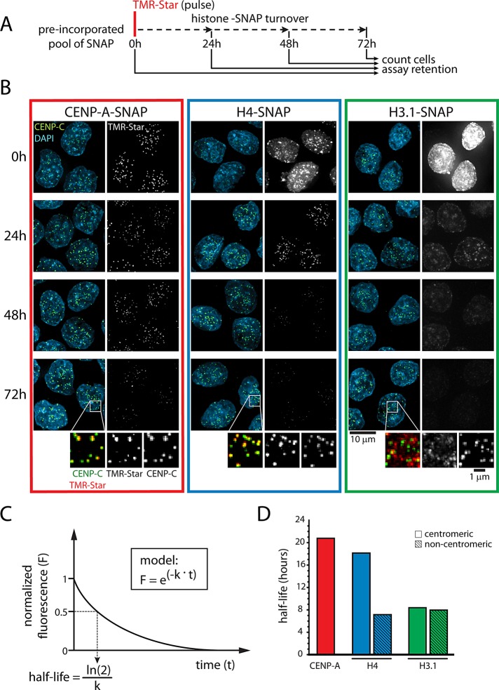
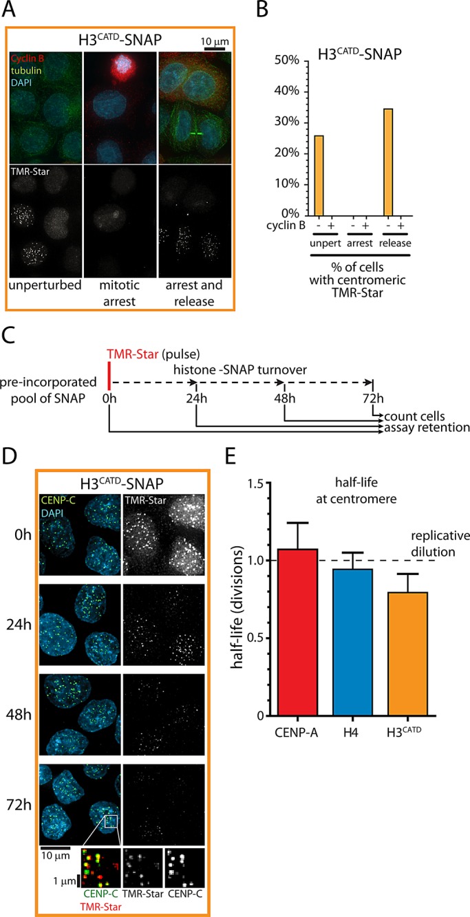
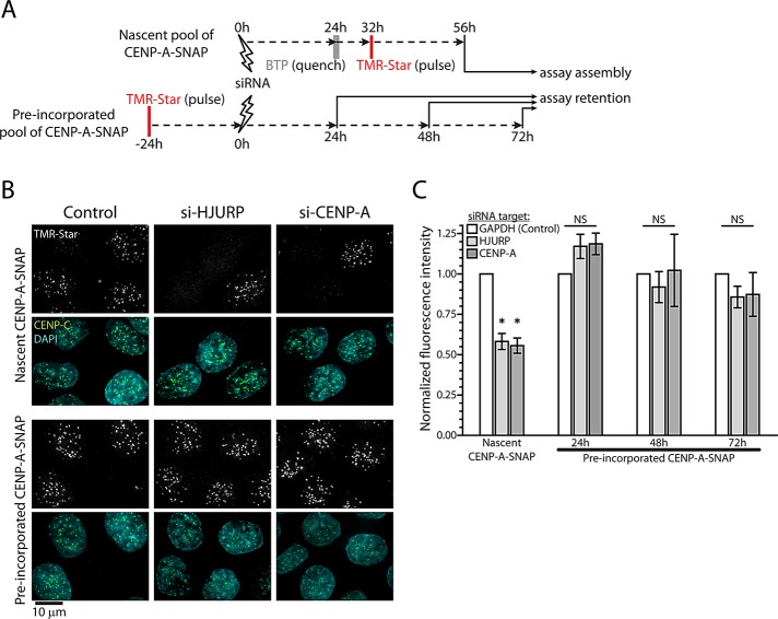
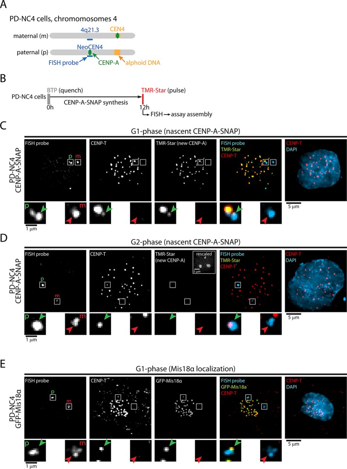
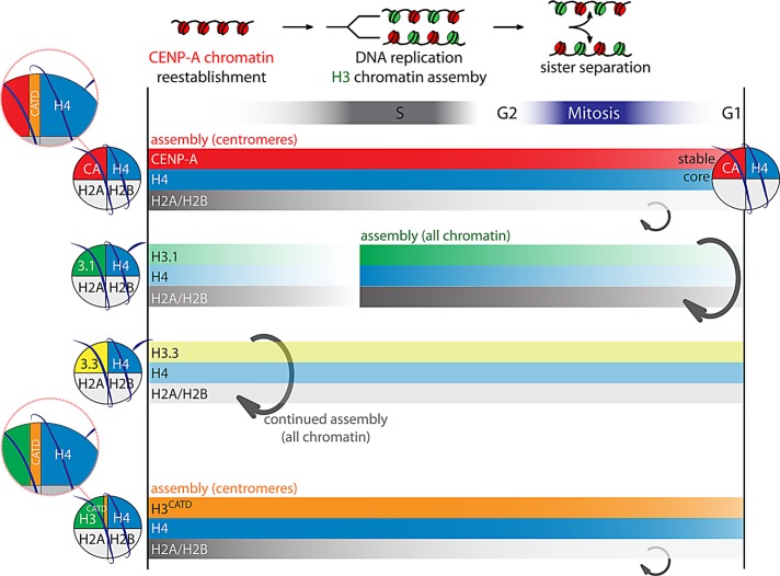
Centromeres are the site of kinetochore formation during mitosis. Centromere protein A (CENP-A), the centromere-specific histone H3 variant, is essential for the epigenetic maintenance of centromere position. Previously we showed that newly synthesized CENP-A is targeted to centromeres exclusively during early G1 phase and is subsequently maintained across mitotic divisions. Using SNAP-based fluorescent pulse labeling, we now demonstrate that cell cycle-restricted chromatin assembly at centromeres is unique to CENP-A nucleosomes and does not involve assembly of other H3 variants. Strikingly, stable retention is restricted to the CENP-A/H4 core of the nucleosome, which we find to outlast general chromatin across several cell divisions. We further show that cell cycle timing of CENP-A assembly is independent of centromeric DNA sequences and instead is mediated by the CENP-A targeting domain. Unexpectedly, this domain also induces stable transmission of centromeric nucleosomes, independent of the CENP-A deposition factor HJURP. This demonstrates that intrinsic properties of the CENP-A protein direct its cell cycle-restricted assembly and induces quantitative mitotic transmission of the CENP-A/H4 nucleosome core, ensuring long-term stability and epigenetic maintenance of centromere position.


Figures

FIGURE 1:
FIGURE 1:
H4, but not H3.1, H3.3, or H2B, is coassembled with CENP-A in G1 phase. (A) Outline of quench-chase-pulse labeling strategy, allowing visualization of a newly synthesized pool of SNAP, followed by Triton-based preextraction. (B) Results of A for indicated histone–SNAP fusion proteins. Enlargement to the right shows rescaled images to indicate colocalization of newly synthesized H4-SNAP with centromeres (marked by CENP-C). Enlargements below show single–focal plane images to indicate specific subnuclear assembly patterns. Blue, green, and red arrows show G1, early S, and mid/late S phase cells, respectively. (C) Outline of quench-chase-pulse experiment on synchronized cells. (D) Results of C for SNAP-tagged histone proteins. CENP-C staining indicates centromere positions. Enlargement shows colocalization of newly synthesized H4-SNAP with centromeres.
FIGURE 2:
FIGURE 2:
Assembly of CENP-A and H4 depends on passage through mitosis. (A) Outline of quench-chase-pulse experiment in unperturbed cells, combined with nocodazole treatment, or with nocodazole treatment and washout. (B) Results of A for CENP-A–SNAP and H4-SNAP. Cyclin B and tubulin staining indicate G2 and G1 (midbodies) status, respectively. (C) Quantification of D. Approximately 200–300 cells were analyzed for each condition. Note that during the 8-h chase, cells transit through ∼40% of an ∼22 h cell cycle, indicating the maximum expected percentage of cells entering G1 phase.
FIGURE 3:
FIGURE 3:
CENP-A and H4 are preferentially maintained at centromeres. (A) Outline of pulse-chase experiment allowing for analysis of a preincorporated pool of SNAP for up to 72 h. At each time point, cells were counted to allow accurate quantification of SNAP turnover per cell division. (B) Results of A for CENP-A–SNAP, H4-SNAP, and H3.1-SNAP. Enlargements show rescaled images of remaining protein pool after 72 h (see also Supplemental Figure S3A). (C) Schematic outline for calculation of histone half-life in D and E (see Materials and Methods). (D) Half-life measurements of centromeric and noncentromeric histone pools as a function of time from experiment in B. Noncentromeric CENP-A is below detection and therefore not measured. Data are obtained from between 570 and 1464 (centromeric) foci for each time point.
FIGURE 4:
FIGURE 4:
CATD determines G1 phase assembly and stable transmission of CENP-A nucleosomes. (A) Results of experiment as outlined in Figure 2A for H3CATD-SNAP. Cyclin B and tubulin staining indicate G2 and G1 (midbodies) status, respectively. (B) Quantification of A. Approximately 200–300 cells were analyzed for each condition. Note that during the 8-h chase, cells transit through ∼40% of an ∼22 h cell cycle, indicating the maximum expected percentage of cells entering G1 phase. (C) Outline of pulse-chase experiment analogous to experiment in Figure 3. At each time point, cells were counted to allow accurate quantification of SNAP turnover per cell division. (D) Results of C. Enlargements show rescaled images of remaining protein pool after 72 h (see also Supplemental Figure S3A). (E) Determination of centromeric histone half-life as a function of population doublings from experiments shown in Figures 3B and 4D. Dashed line indicates expected values for proteins that are never lost but merely redistributed as cells divide. Average and SEM of three independent experiments.
FIGURE 5:
FIGURE 5:
HJURP is dispensable for stable retention of CENP-A. (A) Outline of quench-chase-pulse and pulse-chase experiment combined with siRNA-mediated protein depletion. Note that quench-chase-pulse and pulse-chase experiments were done in parallel to minimize variation of RNAi efficiency. (B) Results of A after depletion of GAPDH (control), HJURP, or CENP-A. Images are displayed for nascent CENP-A-SNAP and the preincorporated pool 24 h after target protein depletion. (C) Quantification of centromeric TMR-Star fluorescence after depletion of GAPDH (white), HJURP (light gray), or CENP-A (dark gray) for indicated protein pools of CENP-A–SNAP. Results were normalized against control RNAi. Average and SEM for at least three independent experiments. Asterisks and NS, respectively, indicate statistically significant (p < 0.01) and nonsignificant (p > 0.05) differences from control samples in paired t tests.
FIGURE 6:
FIGURE 6:
Timing of CENP-A assembly is maintained at neocentromeres. (A) Cartoon of maternal (canonical centromere) and paternal (neocentric) chromosome 4 in PD-NC4 cells. Indicated is chromosomal position 4q21.3, the site of neocentromere formation and the hybridization site of the FISH probe used. (B) Outline of quench-chase-pulse experiment in CENP-A–SNAP–expressing PD-NC4 cells. (C, D) Results of B for cells in G1 phase (C) or G2 phase (D), as indicated by nucleolar TMR staining, shown in rescaled inset. CENP-T indicates centromere positions. Enlargements display images of the hybridization sites of the FISH probe. Green arrows indicate the neocentromere, and red arrows show the homologous region on the maternal chromosome. (E) GFP-Mis18α–expressing PD-NC4 cells were stained for GFP and for 4q21.3 by FISH to detect Mis18α and the NeoCEN4, respectively. Enlargements as described above. Paternal (neocentric) and maternal 4q21.3 positions are indicated by p and m, respectively, in C–E.
FIGURE 7:
FIGURE 7:
Model depicting unique features of centromeric nucleosomes. Cell cycle dynamics of different types of nucleosomes are indicated. CENP-A nucleosomes are assembled at centromeres in G1 phase, whereas H3.1 and H3.3 nucleosomes are assembled into general chromatin in S phase and throughout the cell cycle, respectively (Ray-Gallet et al., 2011). Neither H3.1 nor H3.3 nucleosomes are preferentially loaded into centromeric chromatin during G1 phase or any other cell cycle stage. Whereas H2A and H2B are dynamic in all types of nucleosomes, the centromeric CENP-A/H4 core is stable at time scales far surpassing the cell division rate. However, H3.1, H3.3, and noncentromeric H4 turn over more rapidly than CENP-A, and no preferential centromeric maintenance of H3.1 or H3.3 is observed. Key to both temporal assembly and stable transmission is the CATD domain of CENP-A, which forms a stable interface with H4 in both CENP-A and H3CATD nucleosomes (Sekulic et al., 2010; Bassett et al., 2012).

References

    1. Ahmad K, Henikoff S. Histone H3 variants specify modes of chromatin assembly. Proc Natl Acad Sci USA. 2002;99(Suppl 4):16477–16484. - PMC - PubMed
    1. Amor DJ, Bentley K, Ryan J, Perry J, Wong L, Slater H, Choo KHA. Human centromere repositioning “in progress.”. Proc Natl Acad Sci USA. 2004;101:6542–6547. - PMC - PubMed
    1. Barnhart MC, Kuich PHJL, Stellfox ME, Ward JA, Bassett EA, Black BE, Foltz DR. HJURP is a CENP-A chromatin assembly factor sufficient to form a functional de novo kinetochore. J Cell Biol. 2011;194:229–243. - PMC - PubMed
    1. Bassett EA, DeNizio J, Barnhart-Dailey MC, Panchenko T, Sekulic N, Rogers DJ, Foltz DR, Black BE. HJURP uses distinct CENP-A surfaces to recognize and to stabilize CENP-A/histone H4 for centromere assembly. Dev Cell. 2012;22:749–762. - PMC - PubMed
    1. Bassett EA, Wood S, Salimian KJ, Ajith S, Foltz DR, Black BE. Epigenetic centromere specification directs Aurora B accumulation but is insufficient to efficiently correct mitotic errors. J Cell Biol. 2010;190:177–185. - PMC - PubMed

Publication types

MeSH terms

LinkOut - more resources