. 2013 Mar;24(6):785-95.
doi: 10.1091/mbc.E12-07-0522. Epub 2013 Jan 30.

The ERdj5-Sel1L complex facilitates cholera toxin retrotranslocation

Affiliations

The ERdj5-Sel1L complex facilitates cholera toxin retrotranslocation

Jeffrey M Williams et al. Mol Biol Cell. 2013 Mar.

Abstract

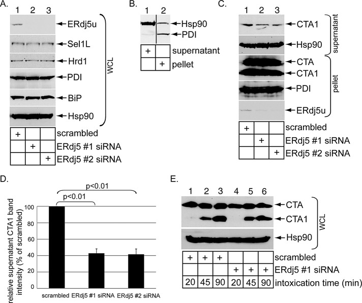
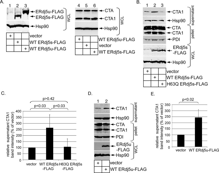
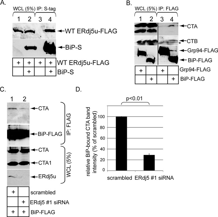
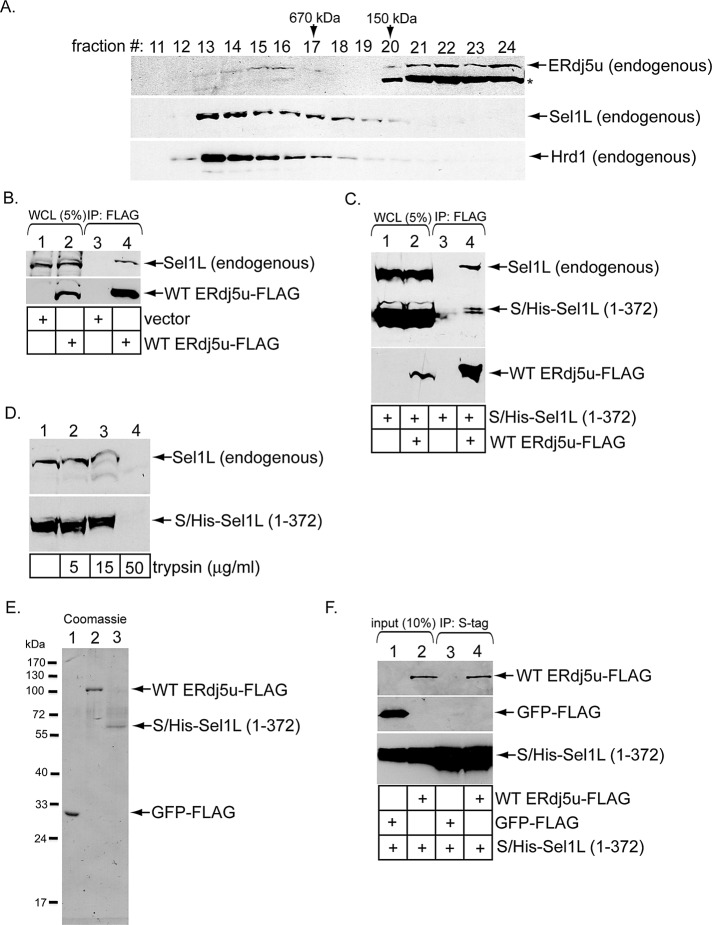
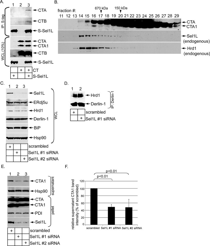
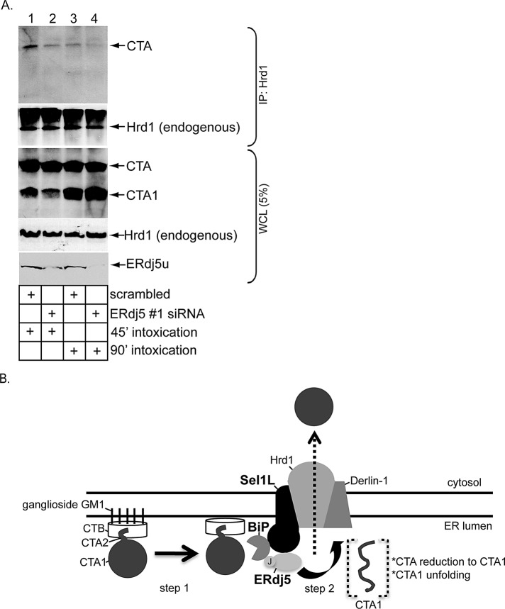
Cholera toxin (CT) traffics from the host cell surface to the endoplasmic reticulum (ER), where the toxin's catalytic CTA1 subunit retrotranslocates to the cytosol to induce toxicity. In the ER, CT is captured by the E3 ubiquitin ligase Hrd1 via an undefined mechanism to prepare for retrotranslocation. Using loss-of-function and gain-of-function approaches, we demonstrate that the ER-resident factor ERdj5 promotes CTA1 retrotranslocation, in part, via its J domain. This Hsp70 cochaperone regulates binding between CTA and the ER Hsp70 BiP, a chaperone previously implicated in toxin retrotranslocation. Importantly, ERdj5 interacts with the Hrd1 adaptor Sel1L directly through Sel1L's N-terminal lumenal domain, thereby linking ERdj5 to the Hrd1 complex. Sel1L itself also binds CTA and facilitates toxin retrotranslocation. By contrast, EDEM1 and OS-9, two established Sel1L binding partners, do not play significant roles in CTA1 retrotranslocation. Our results thus identify two ER factors that promote ER-to-cytosol transport of CTA1. They also indicate that ERdj5, by binding to Sel1L, triggers BiP-toxin interaction proximal to the Hrd1 complex. We postulate this scenario enables the Hrd1-associated retrotranslocation machinery to capture the toxin efficiently once the toxin is released from BiP.


Figures

FIGURE 1:
FIGURE 1:
ERdj5 knockdown decreases CTA1 retrotranslocation. (A) WCLs from 293T cells transfected with a scrambled, ERdj5 #1, or ERdj5 #2 siRNA were analyzed by SDS–PAGE and immunoblotted with the indicated antibodies. The ERdj5 antibody used was from Santa Cruz Biotechnology. (B) Cells were incubated with digitonin and centrifuged. The resulting supernatant and pellet fractions were analyzed for the presence of the cytosolic Hsp90 and ER-resident PDI markers. This protocol represents the fractionation procedure used in the retrotranslocation assay. The vertical black line indicates splicing of the lanes from the same blot. (C) Cells transfected with a scrambled, ERdj5 #1, or ERdj5 #2 siRNA were incubated with CT (10 nM) for 90 min and subjected to the retrotranslocation assay, as in (B). The supernatant and pellet fractions were analyzed by immunoblotting with the indicated antibodies. (D) The CTA1 band intensity generated in (C) was quantified with ImageJ. Mean of four independent experiments. A two-tailed t test was used. Error bars: ±SD. (E) Cells transfected with a scrambled or ERdj5 #1 siRNA were intoxicated with CT (10 nM) for the indicated time and harvested, and the resulting WCLs were analyzed with nonreducing SDS–PAGE followed by immunoblotting with the indicated antibodies.
FIGURE 2:
FIGURE 2:
ERdj5 overexpression stimulates CTA1 retrotranslocation. (A) WCLs derived from 293T cells transfected with vector, WT ERdj5s-FLAG, or WT ERdj5u-FLAG and intoxicated with CT (10 nM) were analyzed by immunoblotting using the indicated antibodies. (B) Cells transfected with vector, WT ERdj5s-FLAG, or H63Q ERdj5s-FLAG were incubated with CT (10 nM) for 90 min and subjected to the retrotranslocation assay, as in Figure 1C. The supernatant and pellet fractions and the WCLs were analyzed by immunoblotting with the indicated antibodies. (C) The CTA1 band intensity in (B) was quantified as in Figure 1D. Mean of five independent experiments. A two-tailed t test was used. Error bars: ± SD. (D) As in (B), except only vector and WT ERdj5u-FLAG were transfected. (E) The CTA1 band intensity in (D) was analyzed as in (C). Mean of seven independent experiments. A two-tailed t test was used. Error bars: ±SD.
FIGURE 3:
FIGURE 3:
ERdj5 binds to BiP and regulates BiP–CTA interaction. (A) Cells were transfected with WT ERdj5u-FLAG alone or in combination with BiP-S, and lysed in a buffer containing 1% deoxy Big CHAP. The resulting WCLs were incubated with S-antibody–conjugated beads, and the immunoprecipitates were subjected to SDS–PAGE followed by immunoblotting with the indicated antibodies. (B) Cells transfected with the control Grp94-FLAG or BiP-FLAG were incubated with CT (10 nM) for 90 min and lysed in a buffer containing 1% deoxy Big CHAP. The resulting WCLs were incubated with FLAG antibody–conjugated beads, and the immunoprecipitates were subjected to SDS–PAGE followed by immunoblotting with CTA, CTB, or FLAG antibodies. The vertical black line indicates antibody splicing of the lanes from the same blot. The asterisk denotes a nonspecific protein that interacts with the CTB antibody. (C) Cells transfected with BiP-FLAG and cotransfected with either a scrambled or ERdj5 #1 siRNA were incubated with CT (10 nM) for 90 min and lysed in a buffer containing 1% deoxy Big CHAP. The resulting WCLs were incubated with FLAG antibody–conjugated beads, and the immunoprecipitates were subjected to SDS–PAGE followed by immunoblotting with the indicated antibodies. WCL samples were also analyzed by immunoblotting with an antibody against CTA and ERdj5. (D) The CTA1 band intensity in (C) was analyzed as in Figure 1D. Mean of three independent experiments. A two-tailed t test was used. Error bars: ±SD.
FIGURE 4:
FIGURE 4:
ERdj5u binds to Sel1L. (A) A WCL derived from 293T cells was subjected to gel filtration. Individual fractions were subjected to SDS–PAGE and analyzed by immunoblotting using the indicated antibodies. The asterisk denotes ERp72 that cross-reacts with an ERdj5 antibody (Proteintech Group). (B) Cells were transfected with vector or WT ERdj5u-FLAG and lysed in a buffer containing 1% deoxy Big CHAP. The resulting WCLs were incubated with FLAG antibody–conjugated beads, and the immunoprecipitates were subjected to SDS–PAGE followed by immunoblotting with the indicated antibodies. (C) Cells transfected with S/His-Sel1L (1–372) alone or S/His-Sel1L (1–372) and WT ERdj5u-FLAG were lysed in a buffer containing 1% deoxy Big CHAP. The resulting WCLs were processed as in (B). (D) A WCL expressing S/His-Sel1L (1–372) was incubated with the indicated trypsin concentration, subjected to SDS–PAGE, and immunoblotted using an antibody against Sel1L. (E) GFP-FLAG, WT ERdj5u-FLAG, and S/His-Sel1L (1–372) were expressed in 293T cells, and the purified proteins were subjected to SDS–PAGE followed by Coomassie blue staining. Fraction 27 during purification of S/His-Sel1L (1–372) is shown. (F) S/His-Sel1L (1–372) was incubated with either GFP-FLAG or WT ERdj5u-FLAG, and the samples were precipitated using S-antibody–conjugated beads. The immunoprecipitates were subjected to SDS–PAGE followed by immunoblotting with the indicated antibodies. The input samples were also analyzed by immunoblotting with the appropriate antibodies.
FIGURE 5:
FIGURE 5:
Sel1L binds to CT and mediates CTA1 retrotranslocation. (A) Cells transfected with or without S-Sel1L were intoxicated with or without CT (10 nM) for 90 min and lysed in a buffer containing 1% deoxy Big CHAP. The resulting WCLs were incubated with S-antibody–conjugated beads, and the immunoprecipitates were subjected to SDS–PAGE followed by immunoblotting with the indicated antibodies. WCL samples were also analyzed by immunoblotting with the appropriate antibodies. (B) As in Figure 4A, except cells were intoxicated with CT (10 nM), semipermeabilized with 0.01% digitonin, and centrifuged to remove toxin in the cytosol; this was followed by solubilization of the resulting pellet with 1% digitonin. (C) As in Figure 1A, except two different Sel1L-directed siRNAs (Sel1 #1 and #2 siRNAs) were used. (D) Cells transfected with a scrambled or Sel1L #1 siRNA were harvested and lysed in a buffer containing 1% deoxy Big CHAP, and the resulting WCLs were subjected to immunoprecipitation with a Derlin-1−specific antibody. The immunoprecipitates were analyzed by immunoblotting using the indicated antibodies. (E) Cells transfected with a scrambled, Sel1L #1 siRNA, or Sel1L #2 siRNA were intoxicated with CT (10 nM) for 90 min. Cells were subjected to the retrotranslocation assay, and the supernatant and pellet fractions were analyzed as in Figure 1C. (F) The CTA1 band intensity in (D) was analyzed as in Figure 1D. Mean of three independent experiments. A two-tailed t test was used. Error bars: ±SD.
FIGURE 6:
FIGURE 6:
ERdj5 knockdown reduces Hrd1–CTA interaction. (A) Cells transfected with either scrambled or ERdj5 #1 siRNA were incubated with CT (10 nM) for 45 min or 90 min and lysed in a buffer containing 1% deoxy Big CHAP. The resulting WCLs were incubated with a Hrd1 antibody, and the immunoprecipitates were subjected to SDS–PAGE followed by immunoblotting with the indicated antibodies. WCL samples were also analyzed by immunoblotting with the indicated antibodies. (B) Model of ERdj5- and Sel1L-dependent CTA1 retrotranslocation. Using its J domain, ERdj5 stimulates BiP's ATPase activity, enabling BiP to engage the toxin (step 1). Because ERdj5 interacts with the Hrd1 adaptor Sel1L, this reaction occurs proximal to the Hrd1-associated complex. On release from BiP, CTA is reduced and unfolded and is competent for retrotranslocation across the Hrd1 membrane complex (step 2). See the text for more detailed discussion.

Similar articles

Cited by

References

    1. Abujarour RJ, Dalal S, Hanson PI, Draper RK. p97 is in a complex with cholera toxin and influences the transport of cholera toxin and related toxins to the cytoplasm. J Biol Chem. 2005;280:15865–15871. - PubMed
    1. Bernardi KM, Forster ML, Lencer WI, Tsai B. Derlin-1 facilitates the retrotranslocation of cholera toxin. Mol Biol Cell. 2008;19:877–884. - PMC - PubMed
    1. Bernardi KM, Williams JM, Kikkert M, van Voorden S, Wiertz EJ, Ye Y, Tsai B. The E3 ubiquitin ligases Hrd1 and gp78 bind to and promote cholera toxin retrotranslocation. Mol Biol Cell. 2010;21:140–151. - PMC - PubMed
    1. Bernasconi R, Galli C, Calanca V, Nakajima T, Molinari M. Stringent requirement for HRD1, SEL1L, OS-9/XTP3-B for disposal of ERAD-LS substrates. J Cell Biol. 2010;188:223–235. - PMC - PubMed
    1. Buck TM, Kolb AR, Boyd CR, Kleyman TR, Brodsky JL. The endoplasmic reticulum-associated degradation of the epithelial sodium channel requires a unique complement of molecular chaperones. Mol Biol Cell. 2010;21:1047–1058. - PMC - PubMed

Publication types

MeSH terms

LinkOut - more resources