. 2013 Apr;24(7):890-900.
doi: 10.1091/mbc.E12-11-0838. Epub 2013 Jan 30.

N-terminal acetylation of the yeast Derlin Der1 is essential for Hrd1 ubiquitin-ligase activity toward luminal ER substrates

Affiliations

N-terminal acetylation of the yeast Derlin Der1 is essential for Hrd1 ubiquitin-ligase activity toward luminal ER substrates

Dimitrios Zattas et al. Mol Biol Cell. 2013 Apr.

Abstract

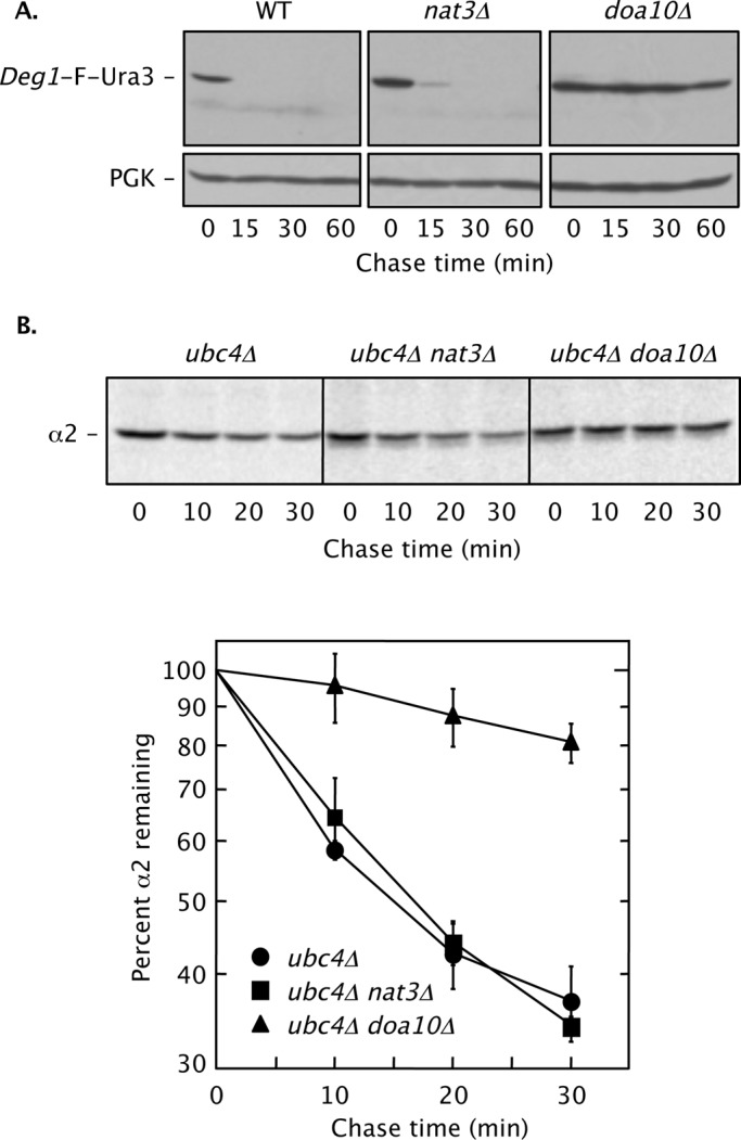
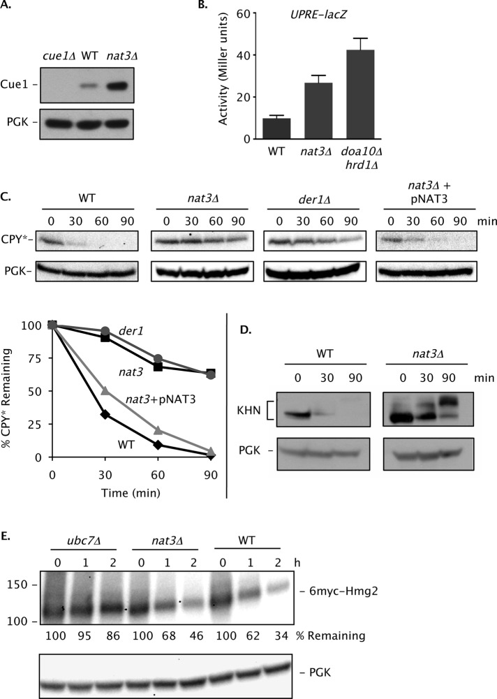
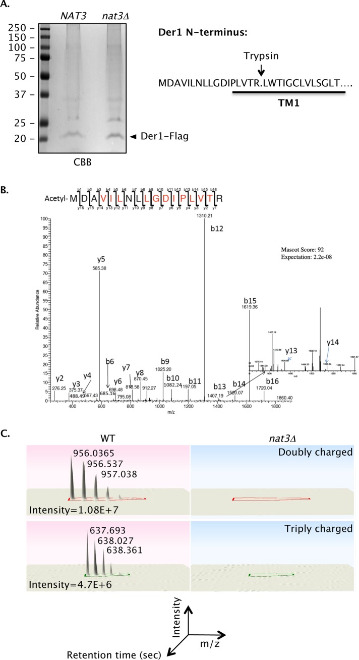
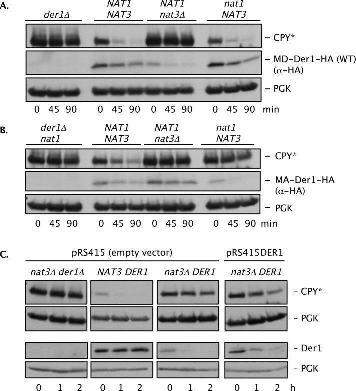
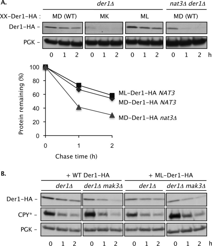
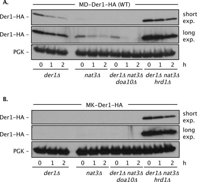
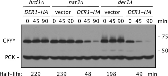
Two conserved ubiquitin ligases, Hrd1 and Doa10, mediate most endoplasmic reticulum-associated protein degradation (ERAD) in yeast. Degradation signals (degrons) recognized by these ubiquitin ligases remain poorly characterized. Doa10 recognizes the Deg1 degron from the MATα2 transcription factor. We previously found that deletion of the gene (NAT3) encoding the catalytic subunit of the NatB N-terminal acetyltransferase weakly stabilized a Deg1-fusion protein. By contrast, a recent analysis of several MATα2 derivatives suggested that N-terminal acetylation of these proteins by NatB was crucial for recognition by Doa10. We now analyze endogenous MATα2 degradation in cells lacking NatB and observe minimal perturbation relative to wild-type cells. However, NatB mutation strongly impairs degradation of ER-luminal Hrd1 substrates. This unexpected defect derives from a failure of Der1, a Hrd1 complex subunit, to be N-terminally acetylated in NatB mutant yeast. We retargeted Der1 to another acetyltransferase to show that it is the only ERAD factor requiring N-terminal acetylation. Preventing Der1 acetylation stimulates its proteolysis via the Hrd1 pathway, at least partially accounting for the ERAD defect observed in the absence of NatB. These results reveal an important role for N-terminal acetylation in controlling Hrd1 ligase activity toward a specific class of ERAD substrates.


Figures

FIGURE 1:
FIGURE 1:
NatB is not essential for Doa10-dependent MATα2 degradation. (A) Cycloheximide chase/immunoblot analysis of Deg1-F-Ura3 degradation in WT (BY4741), nat3Δ (MHY7428), and doa10Δ (MHY3033) strains. Anti-FLAG (F) antibody was used for immunoblotting. Cell culture and incubation after cycloheximide addition were both conducted at 30°C. Deg1-F-Ura3 was expressed from the plasmid p415MET25-Deg1-FL-URA3. PGK, phosphoglycerate kinase (used as a loading control). (B) Pulse-chase analysis of native MATα2 degradation. Cell extracts were immunoprecipitated with anti-MATα2 antibody. Top, representative pulse-chase autoradiogram. Bottom, quantification of degradation kinetics. Each curve represents the average of four independent experiments. Error bars depict standard errors. Yeast strains used: ubc4Δ (MHY498), ubc4Δ nat3Δ (MHY6850), and ubc4Δ doa10Δ (MHY1648).
FIGURE 2:
FIGURE 2:
NatB activity is important for degradation of CPY*, a Hrd1 ERAD substrate. (A) Loss of NatB causes increased levels of the Ubc7 cofactor Cue1 based on anti-Cue1 immunoblotting. PGK, loading control. (B) A mild UPR in cells lacking NatB. WT (MHY501), nat3Δ (MH6599), and doa10Δ hrd1Δ (MHY1703) strains were transformed with the pSZ1 UPRE-lacZ reporter plasmid (Travers et al., 2000), and at least three independent transformants were evaluated for β-galactosidase activity. Error bars represent SDs. (C) Cycloheximide-chase/anti-CPY immunoblot analysis of CPY* in prc1-1 (MHY1366), prc1-1 nat3Δ (MHY6920), and prc1-1 der1Δ (MHY7110) cells. PGK, loading control. Graph represents quantification of the cycloheximide-chase analyses at the top. CPY* levels were normalized to PGK at each time point. (D) KHN, another Hrd1-dependent luminal ER substrate, is also stabilized by loss of Nat3. Degradation was evaluated by cycloheximide-chase/anti-HA immunoblot analysis. (E) Turnover of 6myc-Hmg2, a membrane substrate of Hrd1, does not require NatB. Cycloheximide-chase was followed by anti-myc immunoblot analysis. Percentage of 6myc-Hmg2 remaining was normalized to PGK at each time point. Strains used were MHY7719 (WT), MHY7720 (nat3Δ), and MHY1661 (ubc7Δ).
FIGURE 3:
FIGURE 3:
Nα-acetylation of Der1 by NatB (Nat3/Mdm20). (A) Purified Der1-FLAG from hrd1Δ NAT3 (MHY3032) and hrd1Δ nat3Δ (MH7430) cells. Indicated bands from the Coomassie brilliant blue (CBB)–stained gel were excised, subjected to in-gel trypsin digestion, and evaluated by LC-MS/MS. (B) MS/MS sequencing of the N-terminal tryptic peptide of Der1 isolated from NAT3 (WT). Indicated in red are the y and b ions that matched the sequence of the fragmented N-terminal peptide. MS/MS spectra were searched using the Mascot algorithm. No Nα-acetylated peptide was identified by MS/MS for Der1-FLAG purified from the hrd1Δ nat3Δ (MHY 7430) strain (Table 1). (C) Normalized ion intensity peaks of the acetylated peptide are shown at the bottom using Progenesis LC-MS software (Nonlinear Dynamics, Durham, NC).
FIGURE 4:
FIGURE 4:
N-terminal mutation of Der1 creates a novel Nα-acetyltransferase dependence. (A) WT Der1 (MD-Der1-HA) is sensitive to loss of NatB (Nat3/Mdm20) but not NatA (Ard1/Nat1). CPY* and Der1-HA degradation were measured by cycloheximide chase/immunoblotting in cells with the indicated genotypes (all strains were prc1-1 der1Δ and carried either the pRS314 plasmid [der1Δ] or p414DER1-HA plasmid). PGK, loading control. (B) Conversion of Der1 into a NatA substrate makes ERAD-L sensitive to loss of NatA. The MA-Der1 protein was functional in WT but not nat1 (or nat3Δ) cells. CPY* and MA-Der1-HA degradation was measured in cells with the indicated genotypes (all strains were prc1–1 der1Δ and had either the pRS314 plasmid [der1Δ nat1] or p414der1-D2A-HA plasmid). PGK, loading control. (C) Degradation of CPY* and endogenous Der1 measured by cycloheximide chase/immunoblot analysis in WT and nat3Δ cells. The same extracts were used for anti-CPY and anti-Der1 immunoblotting but were resolved on separate gels. pRS415DER1 is a low-copy (CEN) vector expressing DER1 from its own promoter. PGK, loading control.
FIGURE 5:
FIGURE 5:
Der1 function and degradation rate is modulated by its N-terminal sequence. (A) A C-terminally HA-tagged WT Der1 that begins with the sequence Met-Asp (MD) is degraded slowly in NAT3 cells but is destabilized ∼2- to 2.5-fold in a nat3Δ strain. A mutant Der1 (MK) with a Lys at the second position is strongly destabilized (the anti-HA blot required prolonged exposure compared with the others), whereas the ML mutant is degraded at the same slow rate as WT Der1 (bottom). Der1 amounts for each time point in the plot were normalized to PGK levels. (B) The ML-Der1-HA protein is functional in ERAD-L even when not acetylated (mak3Δ cells). A low-copy plasmid expressing ML-Der1-HA from the DER1 promoter was transformed into cells of the indicated genotypes. PGK, loading control.
FIGURE 6:
FIGURE 6:
Nα-acetylation of Der1 regulates its degradation by the Hrd1 ligase. Degradation of WT MD-Der1 (A) and N-terminally mutated MK-Der1 (B) depends on Hrd1. Degradation of Der1-HA was followed by cycloheximide-chase assay and anti-HA immunoblotting. PGK, loading control. Strains used: prc1–1 der1Δ (MHY7110); prc1–1 der1Δ nat3Δ (MHY7111); der1Δ nat3Δ doa10Δ (MHY7834); and der1Δ nat3Δ hrd1Δ (MHY7839).
FIGURE 7:
FIGURE 7:
Increased Der1-HA levels suppress the CPY* degradation defect in nat3Δ cells. Cells all carried the prc1-1 allele and had the indicated additional chromosomal deletions; the strains were transformed with p415GPD-DER1-HA. The estimated CPY* half-lives were determined by quantitative immunoblotting using antibodies to HA and PGK and normalization to PGK levels. Strains used: prc1–1 hrd1Δ (MHY6855); prc1–1 nat3Δ (MHY6920); and prc1–1 der1Δ (MHY7110).

Similar articles

Cited by

References

    1. Arendt CS, Hochstrasser M. Identification of the yeast 20S proteasome catalytic centers and subunit interactions required for active-site formation. Proc Natl Acad Sci USA. 1997;94:7156–7161. - PMC - PubMed
    1. Bays NW, Gardner RG, Seelig LP, Joazeiro CA, Hampton RY. Hrd1p/Der3p is a membrane-anchored ubiquitin ligase required for ER- associated degradation. Nat Cell Biol. 2001;3:24–29. - PubMed
    1. Biederer T, Volkwein C, Sommer T. Role of Cue1p in ubiquitination and degradation at the ER surface. Science. 1997;278:1806–1809. - PubMed
    1. Carvalho P, Goder V, Rapoport TA. Distinct ubiquitin-ligase complexes define convergent pathways for the degradation of ER proteins. Cell. 2006;126:361–373. - PubMed
    1. Chen P, Johnson P, Sommer T, Jentsch S, Hochstrasser M. Multiple ubiquitin-conjugating enzymes participate in the in vivo degradation of the yeast MATα2 repressor. Cell. 1993;74:357–369. - PubMed

Publication types

MeSH terms