. 2013 Feb 15;19(4):798-808.
doi: 10.1158/1078-0432.CCR-12-1517. Epub 2013 Jan 30.

SHP2 is overexpressed and inhibits pSTAT1-mediated APM component expression, T-cell attracting chemokine secretion, and CTL recognition in head and neck cancer cells

Affiliations

SHP2 is overexpressed and inhibits pSTAT1-mediated APM component expression, T-cell attracting chemokine secretion, and CTL recognition in head and neck cancer cells

Michael S Leibowitz et al. Clin Cancer Res. .

Abstract

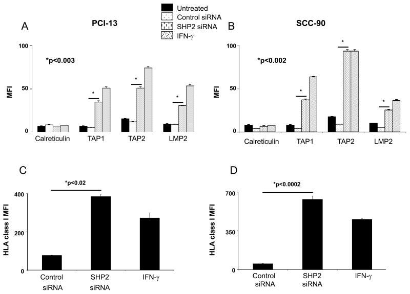
Purpose: Human leukocyte antigen (HLA) class I antigen processing machinery (APM) component downregulation permits escape of malignant cells from recognition by cytotoxic T lymphocytes (CTL) and correlates with poor prognosis in patients with head and neck cancer (HNC). Activated STAT1 (pSTAT1) is necessary for APM component expression in HNC cells. We investigated whether an overexpressed phosphatase was responsible for basal suppression of pSTAT1 and subsequent APM component-mediated immune escape in HNC cells.

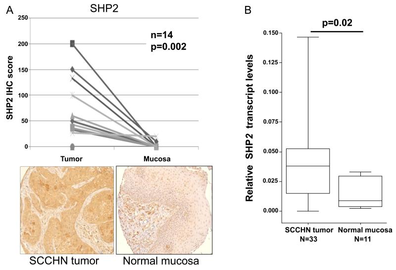
Experimental design: Immunohistochemical staining and reverse transcription PCR of paired HNC tumors was performed for the phosphatases src homology domain-containing phosphatase (SHP)-1 and SHP2. Depletion of phosphatase activity in HNC and STAT1(-/-) tumor cells was achieved by siRNA knockdown. HLA class I-restricted, tumor antigen-specific CTL were used in IFN-γ ELISPOT assays against HNC cells. Chemokine secretion was measured after SHP2 depletion in HNC cells.

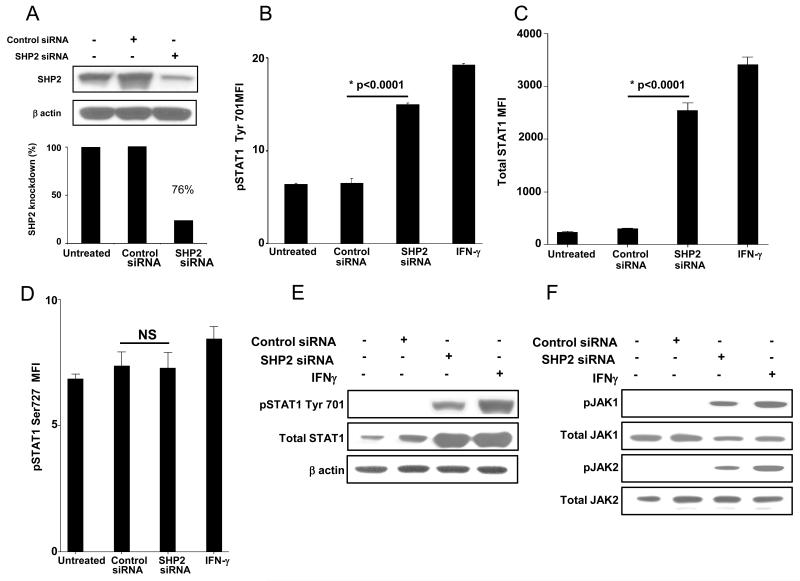
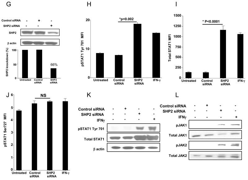
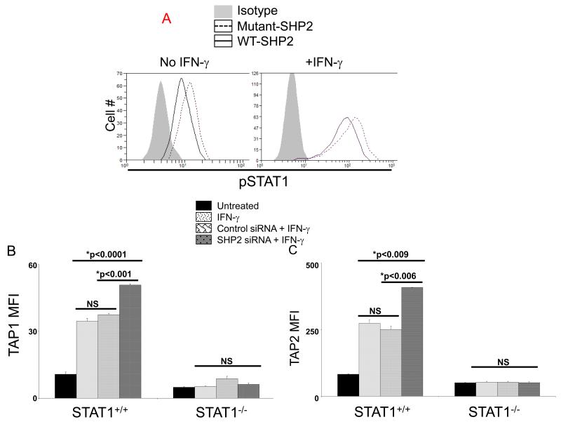
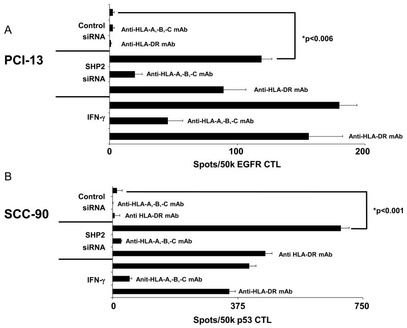
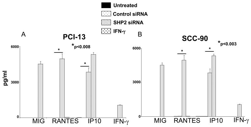
Results: SHP2, but not SHP1, was significantly upregulated in HNC tissues. In HNC cells, SHP2 depletion significantly upregulated expression of pSTAT1 and HLA class I APM components. Overexpression of SHP2 in nonmalignant keratinocytes inhibited IFN-γ-mediated STAT1 phosphorylation, and SHP2 depletion in STAT1(-/-) tumor cells did not significantly induce IFN-γ-mediated APM component expression, verifying STAT1 dependence of SHP2 activity. SHP2 depletion induced recognition of HNC cells by HLA class I-restricted CTL and secretion of inflammatory, T-cell attracting chemokines, RANTES and IP10.

Conclusion: These findings suggest for the first time an important role for SHP2 in APM-mediated escape of HNC cells from CTL recognition. Targeting SHP2 could enhance T-cell-based cancer immunotherapy.


Figures

Figure 1
Figure 1. SHP2 is overexpressed in HNC tissue but not in normal mucosa
(A) HNC tumors and normal adjacent mucosa were stained with an anti-SHP2 mAb (as described in “Materials and Methods”). The IHC semi-quantitative score was derived by two independent pathologists, by multiplying the staining intensity (scored as 0 – 3) by the percent of tumor cells stained (0-100%), to the nearest 5%. IHC scores for each core of a specimen were averaged (n=14) and statistically analyzed. Representative examples at 200x magnification of SHP2 from HNC tissue and normal mucosa are provided. (B) cDNA from HNC tumors (n=33) and normal adjacent mucosa (n=11) were subjected to quantitative real time PCR analysis (as described in materials and methods). Relative expression of shp2 transcript to endogenous control gene (β-actin) was calculated using the ΔCT method: relative expression = 2−ΔCT, where ΔCT = CT (shp2)CT (β-actin). Two-tailed unpaired t-test was performed for statistical analysis.
Figure 2
Figure 2. SHP2 depletion upregulates pSTAT1, pJAK-1/2 and total STAT1
(A-F) PCI-13 and (G-L) SCC-90 cells were transfected with SHP2 siRNA (100 nM, 48 hr) which significantly reduced SHP2 expression in HNC cells by western blot analyses. SHP2 depletion also significantly upregulated pSTAT1 and its target gene product STAT1 as determined by intracellular flow cytometry and immunblotting compared to control siRNA in (B, C, E) PCI-13 and (H, I, K) SCC-90 cells. SHP2 depletion also upregulated pJAK1 (Tyr1022/1023) and pJAK2 (Tyr 1007/1008) compared to control siRNA in (F) PCI-13 and (L) SCC-90 cells. The cells were treated with IFN-γ (100 U/ml=1464 pg/ml, 48h) as a positive control for STAT1 phosphorylation. Data represent three independent experiments and error bars indicate standard error.
Figure 3
Figure 3. SHP2 depletion upregulates HLA class I APM components
PCI-13 and SCC-90 cells were untreated, treated with IFN-γ (100 U/ml=1464 pg/ml, 48 hr), transfected with control siRNA (100 nM, 48 h) or SHP2 siRNA (100 nM, 48 hr). Flow cytometric analyses of intracellular stained cells for TAP1, TAP2, and LMP2 APM components in (A) PCI-13 and (B) SCC-90 cells. Calreticulin, a non-IFN-γ inducible APM component was included to control for global changes in protein expression following transfection. Mean fluorescence intensity (MFI) was measured and error bars indicate standard error. Data represent three independent experiments and error bars indicate standard error. (C) PCI-13 and (D) SCC-90 cells were treated as described above, and HLA class I expression was measured by flow cytometeric MFI. Data represent three independent experiments and error bars indicate standard error.
Figure 4
Figure 4. SHP2 decreases basal and IFN-γ induced pSTAT1 and TAP1/2 expression
(A) Keratinocytes transfected with wild-type SHP2 decreased basal and IFN-γ (10 U/ml=146.4pg/ml, 30 min) induced pSTAT1 compared to mutant SHP2. (C) Parental 2fTGH (STAT1+/+) and U3A (STAT1−/−) fibrosarcoma cells were treated with IFN-γ (10 U/ml=146.4 pg/ml) for 48 hr alone or 24 hours after transfection with SHP2 (50 nM) or control siRNA. Flow cytometric analyses were used to determine TAP1 and TAP2 expression and the data were plotted by MFI. Data represent three independent experiments and error bars indicate standard error.
Figure 5
Figure 5. SHP2 knockdown restores HNC cell recognition by TA-specific CTL
IFN-γ ELISPOT assays were performed to detect recognition by TA-specific CTL of (A) PCI-13 and (B) SCC-90 cells following SHP2 knockdown. Cells were untreated, treated with IFN-γ (100 U/ml=1464 pg/ml, 48 hr), control siRNA (100 nM, 48 hr) or SHP2 siRNA (100 nM, 48 hr). EGFR853-861 and p5365-73 specific cytotoxic T lymphocytes (CTL) were used as effector cells. The HLA class I-specific mAb W6/32 and the HLA-DR-specific mAb L243 were used to demonstrate that the recognition of target cells by TA-specific CTL was HLA class I restricted. Data represent a single experiment performed in triplicate and error bars indicate standard error.
Figure 6
Figure 6. SHP2 siRNA induced the secretion of RANTES and IP10 from HNC cells
Multiplex ELISA (Luminex™) assays were performed on supernatants from (A) PCI-13 and (B) SCC-90 cells following transfection with the indicated siRNA (100 nM, 48 hr) or treatment with IFN-γ (100 U/ml=1464 pg/ml, 48 hr). Data represent three independent experiments and error bars indicate standard error.

References

    1. Koebel CM, Vermi W, Swann JB, Zerafa N, Rodig SJ, Old LJ, et al. Adaptive immunity maintains occult cancer in an equilibrium state. Nature. 2007;450:903–907. - PubMed
    1. Strauss L, Bergmann C, Gooding W, Johnson JT, Whiteside TL. The Frequency and Suppressor Function of CD4+CD25highFoxp3+ T Cells in the Circulation of Patients with Squamous Cell Carcinoma of the Head and Neck. Clin Cancer Res. 2007;13:6301–6311. - PubMed
    1. Kuss I, Hathaway B, Ferris RL, Gooding W, Whiteside TL. Decreased absolute counts of T lymphocyte subsets and their relation to disease in squamous cell carcinoma of the head and neck. Clin Cancer Res. 2004;10:3755–3762. - PubMed
    1. Lopez-Albaitero A, Nayak JV, Ogino T, Machandia A, Gooding W, DeLeo AB, et al. Role of antigen-processing machinery in the in vitro resistance of squamous cell carcinoma of the head and neck cells to recognition by CTL. J Immunol. 2006;176:3402–3409. 2006. - PubMed
    1. Ferris RL, Hunt JL, Ferrone S. Human leukocyte antigen (HLA) class I defects in head and neck cancer: molecular mechanisms and clinical significance. Immunol Res. 2005;33:113–133. - PubMed

Publication types

MeSH terms