. 2013 Jan 30;5(170):170ra13.
doi: 10.1126/scitranslmed.3004912.

CD271(+) bone marrow mesenchymal stem cells may provide a niche for dormant Mycobacterium tuberculosis

Affiliations

CD271(+) bone marrow mesenchymal stem cells may provide a niche for dormant Mycobacterium tuberculosis

Bikul Das et al. Sci Transl Med. .

Abstract

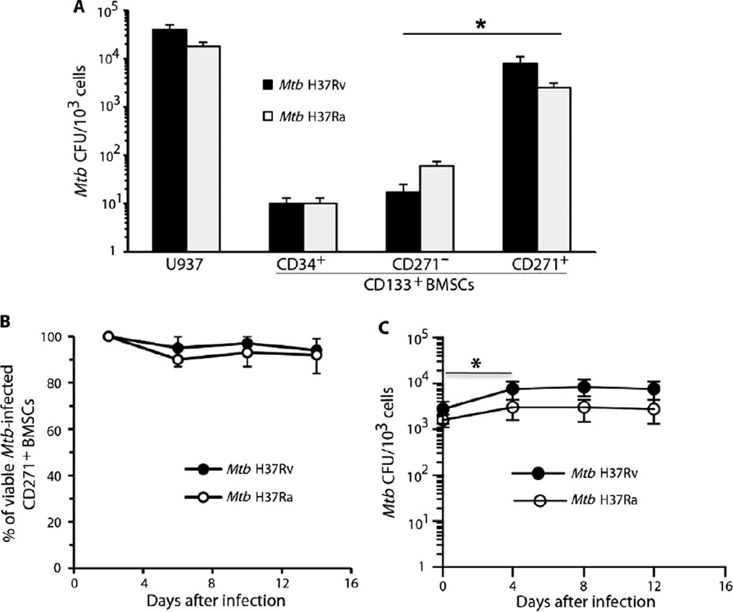
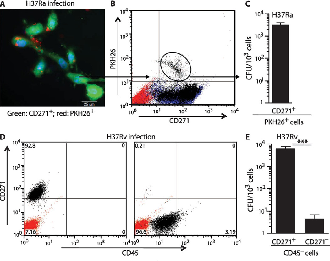
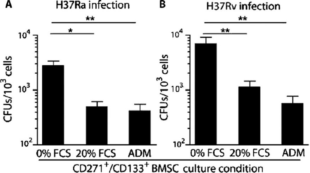
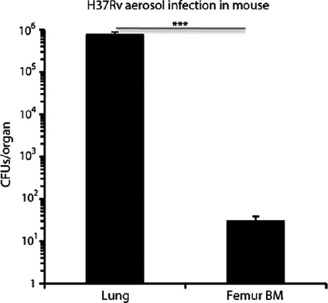
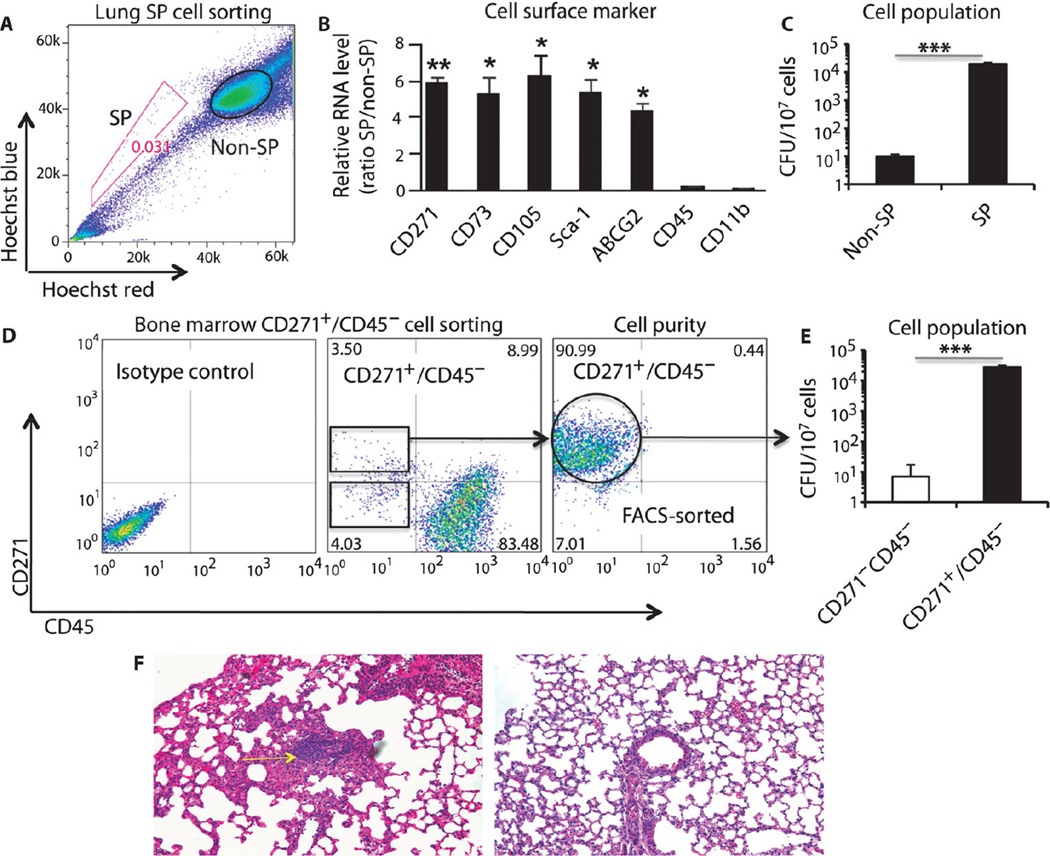
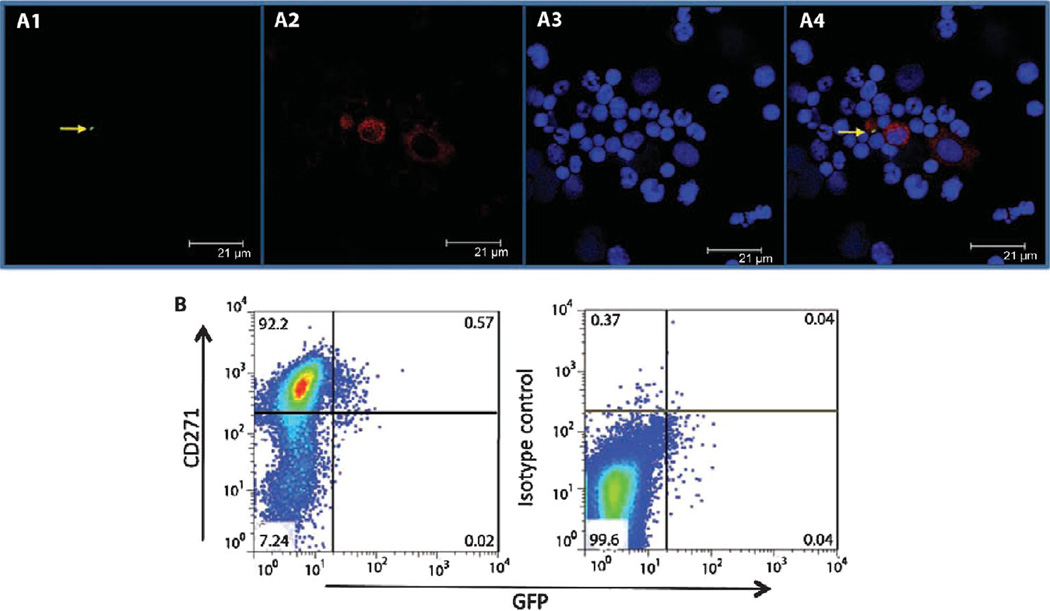
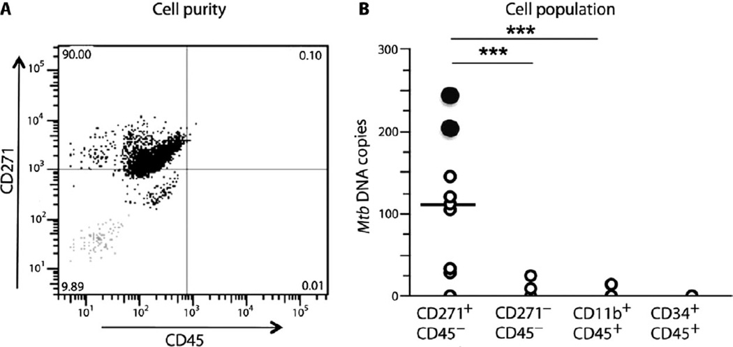
Mycobacterium tuberculosis (Mtb) can persist in hostile intracellular microenvironments evading immune cells and drug treatment. However, the protective cellular niches where Mtb persists remain unclear. We report that Mtb may maintain long-term intracellular viability in a human bone marrow (BM)-derived CD271(+)/CD45(-) mesenchymal stem cell (BM-MSC) population in vitro. We also report that Mtb resides in an equivalent population of BM-MSCs in a mouse model of dormant tuberculosis infection. Viable Mtb was detected in CD271(+)/CD45(-) BM-MSCs isolated from individuals who had successfully completed months of anti-Mtb drug treatment. These results suggest that CD271(+) BM-MSCs may provide a long-term protective intracellular niche in the host in which dormant Mtb can reside.


Figures

Fig. 1
Fig. 1
Mtb infects human CD271+/CD133+ BMSCs. (A) In vitro infection of human CD271+/CD133+ BMSCs with Mtb H37Ra or Mtb H37Rv mycobacterial strains resulted in highest CFU production in the CD271+ fraction of CD133+ BMSCs. Infection of U937 cells was the control. The cells were infected with Mtb [multiplicity of infection (MOI), 5:1], and CFUs were counted after 4 days of in vitro culture. (B) Mtb infection did not reduce CD271+/CD133+ BMSC viability as revealed by the Alamar blue assay (28). (C) Mtb CFUs derived from infected CD271+/CD133+ BMSCs cultured in serum-free medium for 2 weeks (table S1). *P < 0.05, Student’s t test; n = 4 independent experiments.
Fig. 2
Fig. 2
Mtb resides inside human CD271+/CD133+ BMSCs. (A) Epifluorescence image showing amikacin-treated CD271+/CD133+ BMSCs (green) containing intracellular PKH26-labeled Mtb H37Ra strain (red). (B and C) Flow cytometry analysis of infected CD271+/CD133+ BMSCs, and CFUs of PKH26-labeled Mtb H37Ra bacteria derived from infected CD271+/CD133+ BMSCs. (D) Purity of the magnetically sorted CD271+/CD45 and CD271/CD45 cells obtained from H37Rv-infected CD271+/CD133+ BMSCs. (E) Mtb H37Rv CFUs obtained from both of the cell populations depicted in (D). The CD271+/CD133+ BMSCs were infected with Mtb (MOI, 5:1) and grown for 2 weeks in serum-free medium (table S1) before FACS analysis (B) or immunomagnetic sorting (D). The red-stained population in flow cytometry panels (B and D) represents isotype controls. Before harvesting for the Mtb CFU study, infected cells were treated with amikacin (200 µg/ml) for 3 hours to kill extracellular Mtb. *P < 0.05, Student’s t test; n = 4 independent experiments.
Fig. 3
Fig. 3
Viable Mtb reside in undifferentiated CD271+/CD133+/CD45 BM-MSCs. The PKH26+/CD271+ (Fig. 2B) or CD271+/CD45 BM-MSCs, which were obtained from CD271+/CD133+ BMSCs (Fig. 2D), were cultured in vitro in high-serum medium [fetal calf serum (FCS); table S1] or adipogenic differentiation medium (ADM) and cultured for 2 weeks before harvesting. Shown are the numbers of Mtb CFUs grown from each cell population. *P < 0.05, **P < 0.001, Student’s t test; n = 5 independent experiments.
Fig. 4
Fig. 4
Dissemination of Mtb H37Rv to BM in a murine aerosolized model of infection. BM cells (107 mononuclear cells) obtained from mice (female BALB/c) infected with Mtb H37Rv (~100 CFUs) for 4 weeks contained viable Mtb. Shown are numbers of Mtb CFUs grown from mouse femur BM and from lung as a positive control. P < 0.0001, Student’s t test; n = 5 independent experiments.
Fig. 5
Fig. 5
Recovery of viable nonreplicating Mtb from MSCs of infected mice. (A) Flow cytometry profile of infected lung SP and non-SP cells after staining with Hoechst 33342 dye. SP gatingwas performed with verapamil treatment. (B and C) Expression ofMSC markers (B) and detection of viable Mtb CFUs (C) in the FACS-sorted SP and non-SP cellpopulations from infected lungs. (D) Flow cytometry profiles of BM CD271+/CD45 cells from infectedmice. The far right flow cytometry panel represents the purity of the FACS-sorted CD271+/CD45 mouse BM-MSCs. (E) Viable Mtb CFUs were detected in mouse BM-MSC CD271+/CD45 cells (see fig. S3 for characteristics of the CD271+/CD45 BM-MSCs). (F) Granuloma development in the lungs of mice injected with CD271+ BM-MSCs [see (D)] harboring viable dormant Mtb (infected lung, left panel, yellow arrow; the right panel depicts normal lung tissue). Hematoxylin and eosin section was prepared as described (30, 31). Magnification, ×20. *P < 0.05, **P < 0.001, Student’s t test; n = 3 independent experiments.
Fig. 6
Fig. 6
Mtb strain 18b resides in the CD271+/CD45 BM-MSCs of infected mice. (A) Confocal image of flow cytometry–sorted mouse CD271+ BM-MSCs depicting (A1) GFP-tagged Mtb strain 18b (green; indicated by the yellow arrow), (A2) BM cells stained with anti-CD271 monoclonal antibody (red), and (A3) BM nuclei stained with Draq5, a nuclear staining dye. A4, merged image. Images are representative of a typical experiment (n = 3). (B) FACS analysis of the CD271+ BM-MSC population in (A). Representative data are shown for cells staining double positive for CD271 and GFP. Five independent experiments were performed to obtain the percentage of CD271+/GFP+ cells.
Fig. 7
Fig. 7
Viable Mtb persist in the CD271+/CD45 BM-MSCs of treated TB patients. (A) Representative flow cytometry panel showing the purity of the magnetically sorted CD271+/CD45 cells obtained from fresh human BM from subjects previously treated for pulmonary TB. The gray dots (bottom left square) represent the isotype control. (B) CD271+/CD45 BM-MSCs were obtained from previously treated TB patients (DOTS II) who did not show evidence of pulmonary TB. This cell population is enriched for Mtb DNA in eight of nine subjects. The black circles denote two subjects for which viable Mtb could be cultured from their BM-MSCs. A total of 11 post-therapy TB patients were studied. BM from two subjects was verified for enrichment by magnetic sorting (A). The nine other subjects were examined for Mtb DNA and for viable Mtb mycobacteria. Mtb DNA results were analyzed by Student’s t test; ***P < 0.0001.

Similar articles

Cited by

References

    1. Gomez JE, McKinney JD. M. tuberculosis persistence, latency, and drug tolerance. Tuberculosis. 2004;84:29–44. - PubMed
    1. Young DB, Gideon HP, Wilkinson RJ. Eliminating latent tuberculosis. Trends Microbiol. 2009;17:183–188. - PubMed
    1. Via LE, Lin PL, Ray SM, Carrillo J, Allen SS, Eum SY, Taylor K, Klein E, Manjunatha U, Gonzales J, Lee EG, Park SK, Raleigh JA, Cho SN, McMurray DN, Flynn JL, Barry CE., III Tuberculous granulomas are hypoxic in guinea pigs, rabbits, and nonhuman primates. Infect. Immun. 2008;76:2333–2340. - PMC - PubMed
    1. Wayne LG, Hayes LG. An in vitro model for sequential study of shiftdown of Mycobacterium tuberculosis through two stages of nonreplicating persistence. Infect. Immun. 1996;64:2062–2069. - PMC - PubMed
    1. Filippini P, Iona E, Piccaro G, Peyron P, Neyrolles O, Fattorini L. Activity of drug combinations against dormant Mycobacterium tuberculosis. Antimicrob. Agents Chemother. 2010;54:2712–2715. - PMC - PubMed

Publication types

MeSH terms