. 2013 May 15;304(10):C954-65.
doi: 10.1152/ajpcell.00383.2012. Epub 2013 Jan 30.

The cardiomyocyte molecular clock, regulation of Scn5a, and arrhythmia susceptibility

Affiliations

The cardiomyocyte molecular clock, regulation of Scn5a, and arrhythmia susceptibility

Elizabeth A Schroder et al. Am J Physiol Cell Physiol. .

Abstract

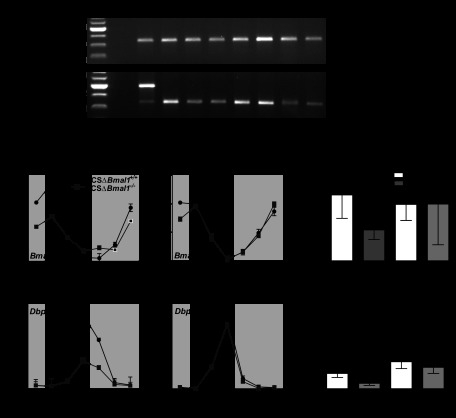
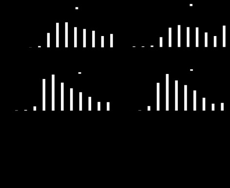
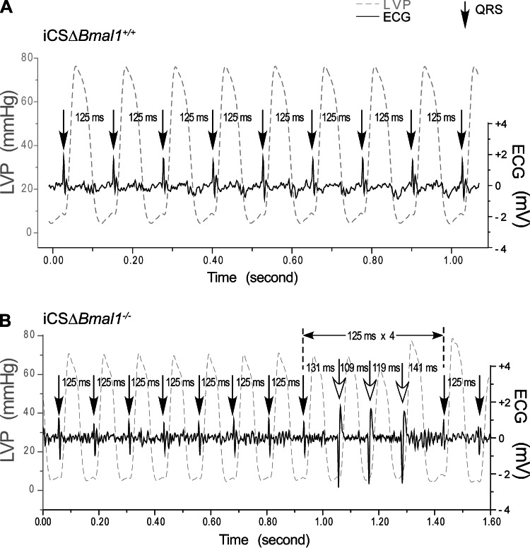
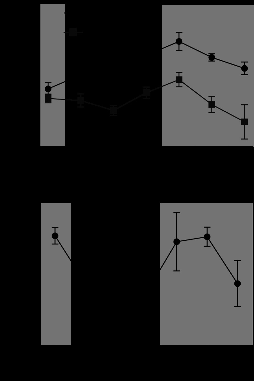
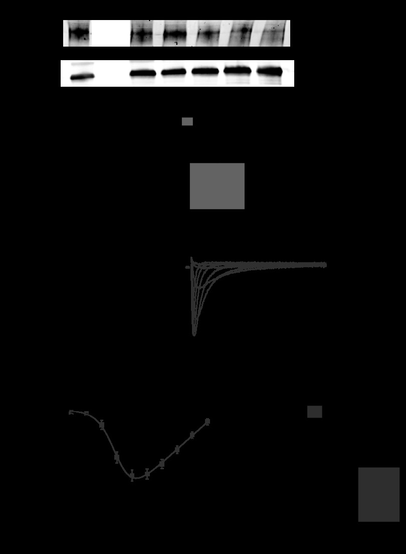
The molecular clock mechanism underlies circadian rhythms and is defined by a transcription-translation feedback loop. Bmal1 encodes a core molecular clock transcription factor. Germline Bmal1 knockout mice show a loss of circadian variation in heart rate and blood pressure, and they develop dilated cardiomyopathy. We tested the role of the molecular clock in adult cardiomyocytes by generating mice that allow for the inducible cardiomyocyte-specific deletion of Bmal1 (iCSΔBmal1). ECG telemetry showed that cardiomyocyte-specific deletion of Bmal1 (iCSΔBmal1(-/-)) in adult mice slowed heart rate, prolonged RR and QRS intervals, and increased episodes of arrhythmia. Moreover, isolated iCSΔBmal1(-/-) hearts were more susceptible to arrhythmia during electromechanical stimulation. Examination of candidate cardiac ion channel genes showed that Scn5a, which encodes the principle cardiac voltage-gated Na(+) channel (Na(V)1.5), was circadianly expressed in control mouse and rat hearts but not in iCSΔBmal1(-/-) hearts. In vitro studies confirmed circadian expression of a human Scn5a promoter-luciferase reporter construct and determined that overexpression of clock factors transactivated the Scn5a promoter. Loss of Scn5a circadian expression in iCSΔBmal1(-/-) hearts was associated with decreased levels of Na(V)1.5 and Na(+) current in ventricular myocytes. We conclude that disruption of the molecular clock in the adult heart slows heart rate, increases arrhythmias, and decreases the functional expression of Scn5a. These findings suggest a potential link between environmental factors that alter the cardiomyocyte molecular clock and factors that influence arrhythmia susceptibility in humans.

Keywords: Na+ current; Scn5a; cardiac excitability; circadian; heart; ion channels.


Figures

Fig. 1.
Fig. 1.
The molecular clock is disrupted in cardiac-specific inducible Bmal1−/− (iCSΔBmal1−/−) mouse hearts. A: representative gels of PCR products showing that recombination at the Bmal1 locus occurs specifically in the heart of the iCSΔBmal1−/− mice but not in other tissues. No recombination was seen in iCSΔBmal1+/+. The recombined gene product shows a band at 571 bp, whereas the nonrecombined product shows a band at 431 bp. Dia, diaphragm; Sol, soleus; Gtn, gastrocnemius; Liv, liver; AA, abdominal aorta; Br, brain. B: the circadian expression profile of Bmal1 and Dbp is significantly blunted specifically in the heart of iCSΔBmal1−/− mice. Dark and light bars represent subjective night and day extrapolated from their prior light:dark (L:D) cycle before release in constant darkness (DD). Bar graphs show average amplitude values with 95% confidence intervals as error bars. n = 4/time point; *P < 0.05.
Fig. 2.
Fig. 2.
Disruption of the molecular clock alters heart rate and QRS intervals. A: 24-h heart rate measured using telemetry implants. Each point represents the half-hour moving average. Note the preservation of circadian rhythmicity and reduction in heart rate for the iCSΔBmal1−/− mice. BPM, beats/min. B and C: average RR and QRS intervals measured from ECG telemetry traces. Note the reduction in heart rate and the trend for longer RR intervals in the iCSΔBmal1−/− mice. *P < 0.05.
Fig. 3.
Fig. 3.
Disruption of the molecular clock alters the diurnal changes in RR and RR variability and increases the incidence of sinoatrial node arrhythmias. A: percentage of beats with the corresponding RR intervals separated by day and night for iCSΔBmal1+/+ and the iCSΔBmal1−/− mice. B: percentage of beats with the corresponding RR intervals over 24 h separated for before (pre-iCSΔBmal1+/+) and after the administration of vehicle or before (pre- iCSΔBmal1−/−) and after disruption of the cardiomyocyte molecular clock. C: representative ECG traces recorded in the pre-iCSΔBmal1−/− and the iCSΔBmal1−/− mice. Mice normally experience isolated pauses, but the iCSΔBmal1−/− mice showed episodes of pauses consistent with sinoatrial node arrhythmias (n = 6 pre-iCSΔBmal1+/+, iCSΔBmal1+/+ and n = 5 pre- iCSΔBmal1−/−, iCSΔBmal1−/−; *P < 0.05).
Fig. 4.
Fig. 4.
Disruption of the cardiomyocyte molecular clock stretch-induced arrhythmias in the isolated heart. Stretch-induced conduction block was tested by increasing preload from 10 mmHg to 12.5, 15, and 20 mmHg in iCSΔBmal1+/+ or iCSΔBmal1−/− hearts paced from the right atrium at 8 Hz. A delay in QRS is due to a delay/block of conduction. A: representative sequence of left ventricular pressure (LVP) and the corresponding ventricular depolarizations (QRS) from an isolated iCSΔBmal1+/+ heart. No conduction block was observed at preloads from 10 to 15 mmHg and only 2 out of 7 hearts showed atrioventricular block at a 20 mmHg preload. B: example of block followed by three wide QRS complexes generated from an ectopic foci (note the inversion of the QRS complex). The three wide QRS complexes following the supraventricular conduction block appeared to be generated from a similar source other than the supraventricular stimulus. The iCSΔBmal1−/− mouse hearts showed conduction block at 12.5 mmHg and became more frequent at 15 mmHg preload (3 out of 6) and 20 mmHg (5 out of 6).
Fig. 5.
Fig. 5.
Scn5a shows a robust circadian oscillation that is lost in iCSΔBmal1−/− mouse hearts. A: the circadian expression profile of Scn5a in the hearts of the iCSΔBmal1+/+ and iCSΔBmal1−/− mice. Dark and light bars represent subjective night and day extrapolated from their prior L:D cycle before release in DD; n = 4/time point. B: Scn5a mRNA is circadian in expression in rat LV; n = 3/time point. WKY, Wistar-Kyoto.
Fig. 6.
Fig. 6.
Bmal1 and CLOCK transactivate the human Scn5a promoter reporter. A: lumicycle data showing the circadian expression pattern of the hScn5a promoter reporter construct transiently transfected in mouse C2C12 (skeletal muscle) cell line; n = 3. B: luciferase assay results of transfection experiments using either the Per1 reporter gene or the hScn5a reporter gene in mouse fibroblast NIH/3T3 cells (n = 6–10/condition). CLOCK (C) and BMAL1 (B) significantly transactivate Per1 and hScn5a reporter genes (black bars) relative to control transfections (white bars). Activation of the hScn5a reporter is significantly diminished when Bmal1R91A was overexpressed with CLOCK (gray bar) or ClockΔ19 was overexpressed with BMAL1 (vertical striped bar). Values are expressed as means ± SE. *P < 0.05 compared with the hScn5a reporter gene; #P < 0.05 compared with hScn5a reporter gene + CLOCK + BMAL.
Fig. 7.
Fig. 7.
Disruption of the cardiomyocyte molecular clock reduces voltage-gated Na+ channel (NaV1.5) levels and Na+ current (INa). A: Western blot data for NaV1.5 in the hearts of iCSΔBmal1+/+ and iCSΔBmal1−/− mice at 58 h in darkness (n = 3/strain; *P < 0.05). B and C: representative families of currents (B) and the mean peak current-voltage (C) relations recorded from iCSΔBmal1+/+ or iCSΔBmal1−/− ventricular cardiomyocytes isolated from mice at 58 h in darkness. The gray and black lines show the robustness of a Boltzmann fit to data. D: iCSΔBmal1−/− ventricular cardiomyocytes had a smaller maximal Na+ conductance (Gmax). *P < 0.05.

Comment in

Similar articles

Cited by

References

    1. CAST II Investigators Effect of the antiarrhythmic agent moricizine on survival after myocardial infarction. The Cardiac Arrhythmia Suppression Trial II Investigators. N Engl J Med 327: 227–233, 1992 - PubMed
    1. CAST Investigators Preliminary report: effect of encainide, and flecainide on mortality in a randomized trial of arrhythmia suppression after myocardial infarction. The Cardiac Arrhythmia Suppression Trial (CAST) Investigators. N Engl J Med 321: 406–412, 1989 - PubMed
    1. Abriel H. Roles and regulation of the cardiac sodium channel Nav1.5: recent insights from experimental studies. Cardiovasc Res 76: 381–389, 2007 - PubMed
    1. Agah R, Frenkel PA, French BA, Michael LH, Overbeek PA, Schneider MD. Gene recombination in postmitotic cells. Targeted expression of Cre recombinase provokes cardiac-restricted, site-specific rearrangement in adult ventricular muscle in vivo. J Clin Invest 100: 169–179, 1997 - PMC - PubMed
    1. Akhtar RA, Reddy AB, Maywood ES, Clayton JD, King VM, Smith AG, Gant TW, Hastings MH, Kyriacou CP. Circadian cycling of the mouse liver transcriptome, as revealed by cDNA microarray, is driven by the suprachiasmatic nucleus. Curr Biol 12: 540–550, 2002 - PubMed

Publication types

MeSH terms

Substances