. 2013 May;62(5):1697-708.
doi: 10.2337/db12-1025. Epub 2013 Jan 30.

Glucagon-like peptide-1 protects against cardiac microvascular injury in diabetes via a cAMP/PKA/Rho-dependent mechanism

Affiliations

Glucagon-like peptide-1 protects against cardiac microvascular injury in diabetes via a cAMP/PKA/Rho-dependent mechanism

Dongjuan Wang et al. Diabetes. 2013 May.

Abstract

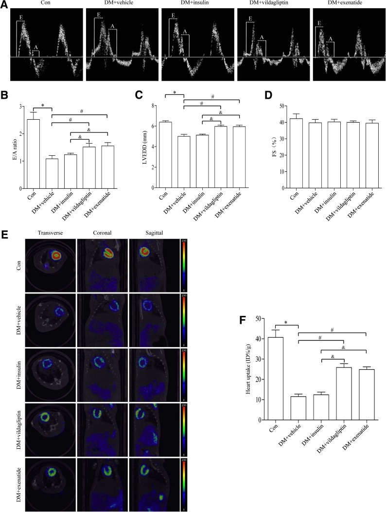
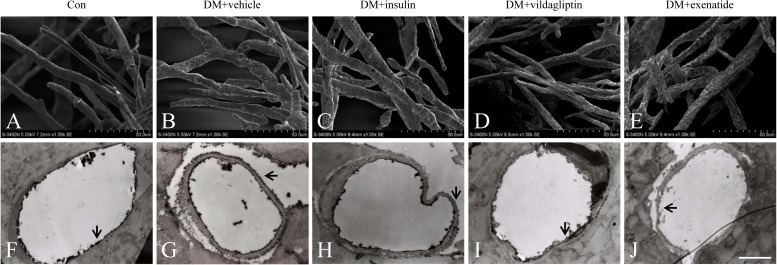
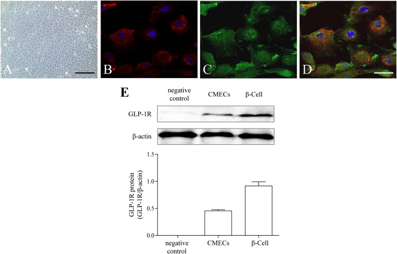
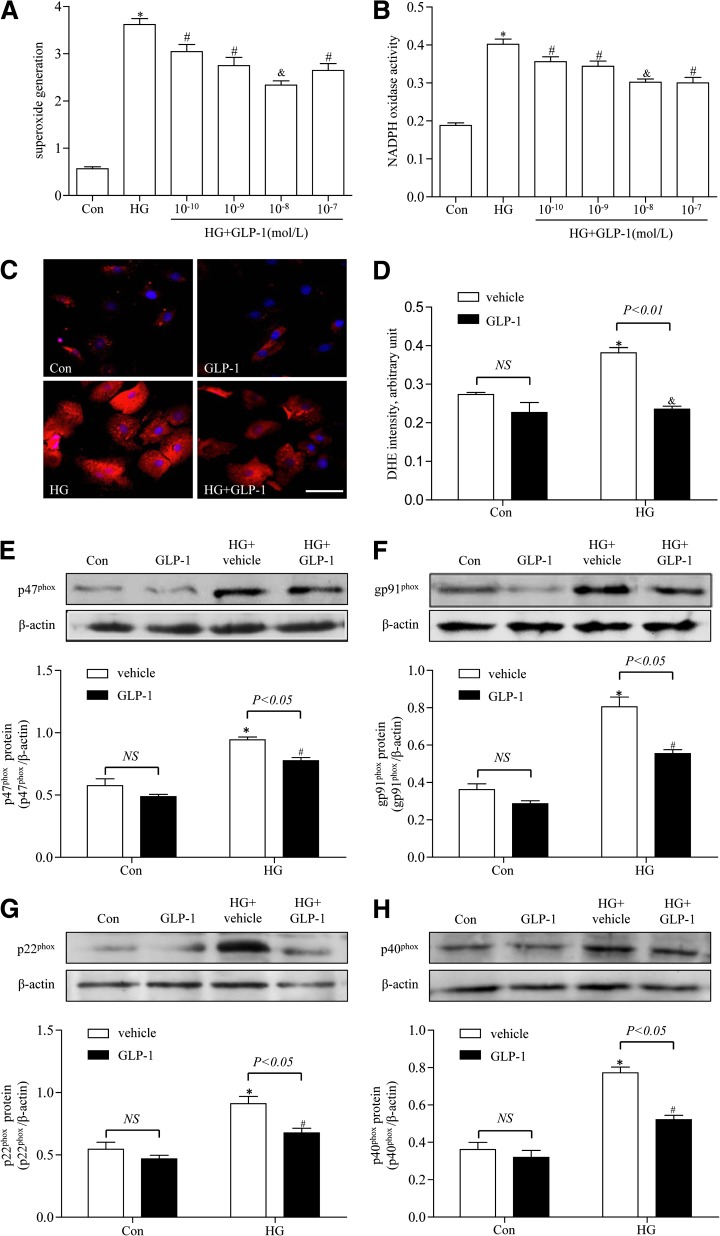
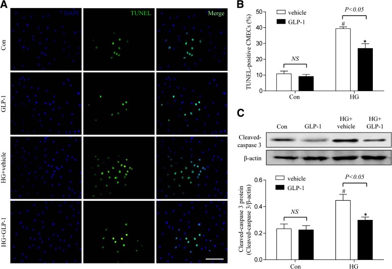
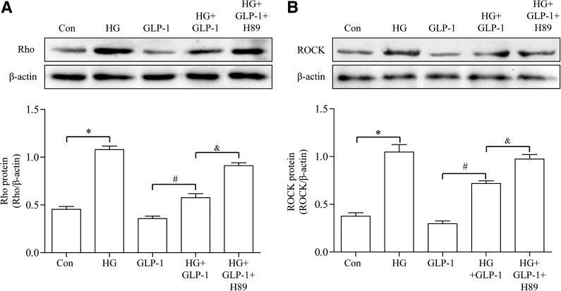
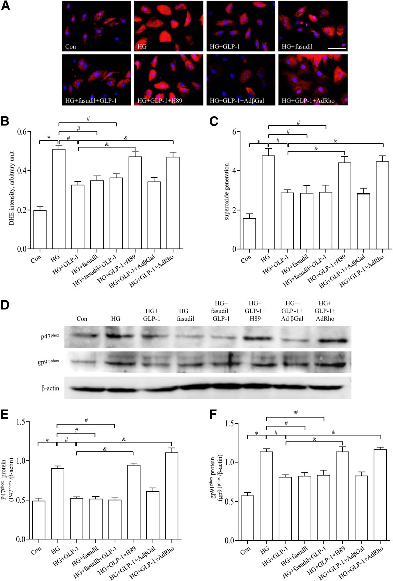
Impaired cardiac microvascular function contributes to cardiovascular complications in diabetes. Glucagon-like peptide-1 (GLP-1) exhibits potential cardioprotective properties in addition to its glucose-lowering effect. This study was designed to evaluate the impact of GLP-1 on cardiac microvascular injury in diabetes and the underlying mechanism involved. Experimental diabetes was induced using streptozotocin in rats. Cohorts of diabetic rats received a 12-week treatment of vildagliptin (dipeptidyl peptidase-4 inhibitor) or exenatide (GLP-1 analog). Experimental diabetes attenuated cardiac function, glucose uptake, and microvascular barrier function, which were significantly improved by vildagliptin or exenatide treatment. Cardiac microvascular endothelial cells (CMECs) were isolated and cultured in normal or high glucose medium with or without GLP-1. GLP-1 decreased high-glucose-induced reactive oxygen species production and apoptotic index, as well as the levels of NADPH oxidase such as p47(phox) and gp91(phox). Furthermore, cAMP/PKA (cAMP-dependent protein kinase activity) was increased and Rho-expression was decreased in high-glucose-induced CMECs after GLP-1 treatment. In conclusion, GLP-1 could protect the cardiac microvessels against oxidative stress, apoptosis, and the resultant microvascular barrier dysfunction in diabetes, which may contribute to the improvement of cardiac function and cardiac glucose metabolism in diabetes. The protective effects of GLP-1 are dependent on downstream inhibition of Rho through a cAMP/PKA-mediated pathway.


Figures

FIG. 1.
FIG. 1.
Effects of vildagliptin or exenatide on cardiac function and cardiac glucose metabolism in diabetes. A: Representative mitral flow patterns from pulsed Doppler. B: Quantification of E/A ratio. C: Measurement of left ventricular end-diastolic diameter after 12 weeks of drug treatment. D: Measurement of fractional shortening (FS) after 12 weeks of drug treatment. E: Representative PET/CT scan images from coronal, sagittal, and transverse view from each group. Higher glucose uptake level is shown as an increase in the intensity of red color as indicated on the color scale bar shown. F: Quantification of accumulated 18F-FDG in the heart. Data are expressed as mean ± SD (n = 6). *P <0.05 vs. control group; #P <0.05 vs. Diabetes + vehicle group; &P <0.05 vs. Diabetes + insulin group. DM, diabetes; E, early mitral diastolic wave; A, late mitral diastolic wave; LVEDD, left ventricular end-diastolic diameter.
FIG. 2.
FIG. 2.
Effects of vildagliptin or exenatide on cardiac microvascular integrity and permeability. A: The surface of cardiac microvessels in control animals was smooth and well-integrated. B: Cardiac microvessels in diabetic animals were visualized to be highly irregular and rough. C: Cardiac microvessels treated with insulin. D and E: Cardiac microvessels with vildagliptin or exenatide treatment exhibited integrity and vessel integration. F: Lanthanum nitrate was regulated to the blood vessel lumen in control animals. G: Lanthanum nitrate was found to diffuse across vessel lumen to the basal lamina in diabetic animals. H: Cardiac microvessels were treated with insulin. I and J: Diffusion of lanthanum nitrate across CMECs was attenuated in vildagliptin or exenatide treatment (scale bar, 2 μm). The extracellular tracer lanthanum nitrate is highlighted with arrows. DM, diabetes.
FIG. 3.
FIG. 3.
Characterization of CMECs and detection of GLP-1R. A: CMEC monolayer presents cobblestone appearance by phase-contrast microscopy (scale bar, 40 μm). B: Uptake of Dil-acetylated low-density lipoprotein (Dil-Ac-LDL) by immunofluorescence (red, Dil-Ac-LDL; blue, DAPI). C: Expression of GLP-1R by immunofluorescence (green, GLP-1R; scale bar, 10 μm). D: Merge between B and C. E: Western blot analysis for GLP-1R. β-Cells were used as positive control.
FIG. 4.
FIG. 4.
Effects of GLP-1 on oxidative stress in high-glucose–induced CMECs. A: GLP-1 inhibits high-glucose–induced superoxide generation in CMECs dose dependently. B: GLP-1 inhibits high-glucose–induced NADPH oxidase activity in CMECs dose dependently. C: Representative images of dihydroethidine (DHE) staining of CMECs (red, DHE; blue, DAPI; scale bar, 25 μm). D: The average fluorescence intensity from five fields was summarized. EH: Western blot assay for p47phox, gp91phox, p22phox, and p40phox protein expression. *P <0.01 vs. control group; #P <0.05 vs. high-glucose (HG) group; &P <0.01 vs. HG group. Con, control.
FIG. 5.
FIG. 5.
GLP-1 exerted antiapoptotic effect on high-glucose–induced CMECs. A: Representative images of immunostaining for apoptotic (TUNEL) cells. Nuclei were labeled with DAPI (scale bar, 25 μm). B: Quantification of apoptotic nuclei by Image-Pro Plus software. C: Western blot assay for cleaved caspase-3 protein expression. *P <0.05 vs. high-glucose (HG) plus vehicle group; #P <0.05 vs. control plus vehicle group. Con, control.
FIG. 6.
FIG. 6.
GLP-1 suppressed high-glucose (HG)-induced activation of Rho. A: Western blot assay for Rho protein expression. B: Western blot assay for ROCK protein expression. *P <0.05 vs. control group; #P <0.05 vs. HG group; &P <0.05 vs. HG plus GLP-1 group. Con, control; H89, PKA inhibitor.
FIG. 7.
FIG. 7.
Effects of GLP-1, fasudil, and H89 on high-glucose (HG)-induced oxidative stress in CMECs. A: Representative diagram showing dihydroethidine (DHE) staining of CMECs (red, DHE; blue, DAPI; scale bar, 25 μm). B: The average fluorescence intensity from five fields was summarized. C: Superoxide generation of CMECs in different groups. D: Western blot assay for p47phox protein and gp91phox protein expression. E and F: Quantitative analysis of p47phox and gp91phox expression. *P <0.01 vs. control group; #P <0.01 vs. HG group; &P <0.05 vs. HG plus GLP-1 group. Con, control; H89, PKA inhibitor.

References

    1. Mazzone T. Intensive glucose lowering and cardiovascular disease prevention in diabetes: reconciling the recent clinical trial data. Circulation 2010;122:2201–2211 - PMC - PubMed
    1. Acar E, Ural D, Bildirici U, Sahin T, Yilmaz I. Diabetic cardiomyopathy. Anadolu Kardiyol Derg 2011;11:732–737 - PubMed
    1. Voulgari C, Papadogiannis D, Tentolouris N. Diabetic cardiomyopathy: from the pathophysiology of the cardiac myocytes to current diagnosis and management strategies. Vasc Health Risk Manag 2010;6:883–903 - PMC - PubMed
    1. Ismail-Beigi F, Craven T, Banerji MA, et al. ACCORD trial group Effect of intensive treatment of hyperglycaemia on microvascular outcomes in type 2 diabetes: an analysis of the ACCORD randomised trial. Lancet 2010;376:419–430 - PMC - PubMed
    1. Hao M, Li SY, Sun CK, et al. Amelioration effects of berberine on diabetic microendothelial injury model by the combination of high glucose and advanced glycation end products in vitro. Eur J Pharmacol 2011;654:320–325 - PubMed

Publication types

MeSH terms