. 2013 Jul;133(7):1827-33.
doi: 10.1038/jid.2013.48. Epub 2013 Jan 30.

The hematopoietic stem cell regulatory gene latexin has tumor-suppressive properties in malignant melanoma

Affiliations

The hematopoietic stem cell regulatory gene latexin has tumor-suppressive properties in malignant melanoma

Viswanathan Muthusamy et al. J Invest Dermatol. 2013 Jul.

Abstract

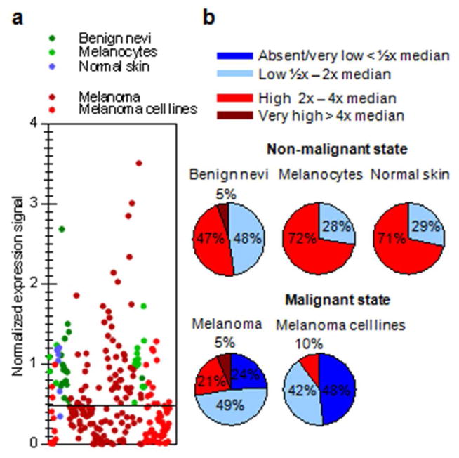
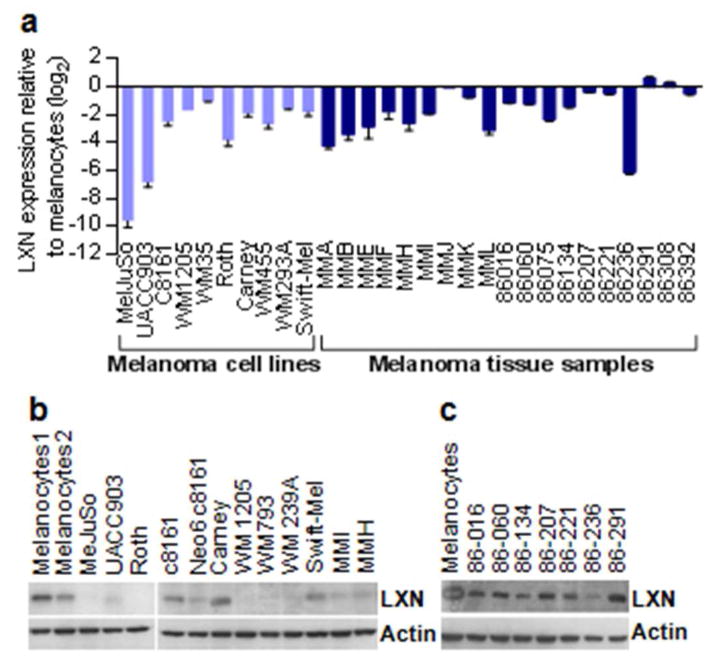
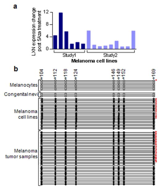
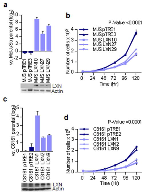
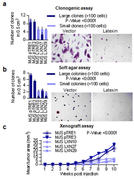
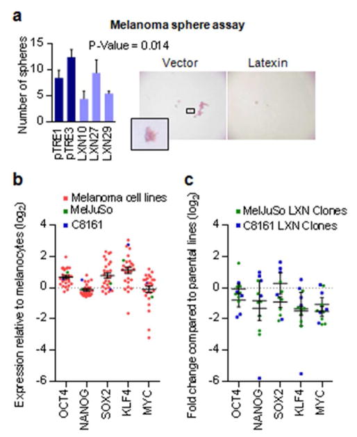
Despite recent advancements in therapy, melanoma remains a highly lethal skin cancer. A better understanding of the genetic and epigenetic changes responsible for melanoma formation and progression could result in the development of more effective treatments. Advanced melanomas are known to exhibit widespread promoter region CpG island methylation leading to the inactivation of key tumor suppressor genes. Meta-analyses of relevant microarray data sets revealed the hematopoietic stem cell regulator gene latexin (LXN) to be commonly downregulated in approximately 50% of melanomas. The CpG island in the promoter region of LXN was almost universally hypermethylated in melanoma cell lines and tumors, and treatment of the cell lines with the demethylating drug 5-aza-2'-deoxycytidine resulted in increased LXN expression. In this paper, we demonstrate that the exogenous expression of LXN in melanoma cell lines results in a significant inhibition of tumor cell proliferation. In addition, we show that the increased expression of LXN in these lines correlates with reduction in the expression levels of stem cell transcription factors OCT4, NANOG, SOX2, KLF4, and MYCN, indicating that LXN may exert its tumor-suppressive function by altering the stem cell-like properties of melanoma cells.


Conflict of interest statement

Conflict of interest

The authors state no conflict of interest

Figures

Figure 1
Figure 1. Expression analysis of LXN in human melanoma and melanocytes
a) Meta-analysis of GEO datasets showing LXN mRNA expression in normal cells – melanocytes, normal skin and benign nevi and malignant cells – melanoma cell lines and tumors. Each dot represents the ratio of expression signal of an individual sample to the median signal of normal samples in that experiment. (b) Stratified levels of LXN expression in normal and malignant states
Figure 2
Figure 2. Validation of reduced LXN expression in melanoma
(a) Quantitiative PCR analysis of LXN expression in melanoma cell lines and tumor tissue samples. (b) Western blot for LXN protein expression in melanoma cell lines (c) Western blot showing LXN protein expression in tumor samples compared to cultured melanocytes
Figure 3
Figure 3. Promoter region hypermethylation leads to silencing of LXN in melanoma
(a) Analysis of microarray data from two previous experiments showing restoration of LXN expression in melanoma cell lines upon treatment with methylation reversing drug 5 Aza 2 deoxycytidine. Study1: light red (Muthusamy et al 2006); Study2: dark red (unpublished data). (b) Sanger bisulfite sequencing of the LXN gene promoter CpG island in melanocytes, melanoma cell lines and tumor samples. CpG positions are indicated by circles in scale to their location in the promoter region. Clear circles indicate absence of methylation, filled circles represent methylated cytosine. The numbers at the top indicate distance from the transcription start site. Stars indicate that the methylation status of these samples were described previously (Muthusamy et al 2006)
Figure 4
Figure 4. Inhibition of cell proliferation by exogenous expression of LXN
(a) Quantitative PCR and western blot analysis of LXN expression in the vector and LXN transfected clones of the MelJuSo melanoma cell line. (b) Growth curves showing differences in proliferation of LXN transfected and vector control lines of MelJuSo. (c) Quantitative PCR and western blot analysis of LXN expression in the vector and LXN transfected clones of C8161 melanoma cell line. (d) Growth curves showing difference in proliferation of LXN transfected and vector control lines of C8161. Note that original LXN protein was expressed in the original parental line which is reflected in the vector transfected controls
Figure 5
Figure 5. Tumor suppressive properties of LXN demonstrated in LXN negative melanoma cell line
(a) In vitro colony formation assay in LXN transfected MelJuSo compared to vector controls. Large clones comprising >100 cells are represented by dark shading while small clones comprising <100 cells are represented by lighter shading. The results are an average of counts from 10 random squares using a scoring grid, with 5 mm2 squares (b) In vitro colony formation in soft agar by LXN transfected MelJuSo compared to vector controls. Large clones comprising >100 cells are represented by dark shading while small clones comprising <100 cells are represented by lighter shading. The results are an average of counts from 10 random squares using a scoring grid, with 5 mm2 squares (c) In vivo xenograft assay showing reduced formation capability of LXN transfected MelJuSo compared to vector controls. The results depict tumors formed at a total of four injection sites in two mice per condition.
Figure 6
Figure 6. Changes in tumor progenitor properties upon reexpression of LXN
(a) In vitro melanoma sphere formation assay in non-adherent conditions showing reduced numbers of spheres in LXN-transfected clones compared to vector (pTRE) transfected controls. Data shown is for a seeding of 1000 cells. The results represent an average of two replicate experiments. (b) Expression of stem cell transcription factors in melanoma cell lines compared to melanocytes derived from microarray expression data. (c) Expression of stem cell transcription factors in the LXN expressing clones of MelJuSo and C8161 compared to parental lines.

Similar articles

Cited by

References

    1. Aagaard A, Listwan P, Cowieson N, Huber T, Ravasi T, Wells CA, et al. An inflammatory role for the mammalian carboxypeptidase inhibitor latexin: relationship to cystatins and the tumor suppressor TIG1. Structure. 2005;13:309–17. - PubMed
    1. Aitken J, Welch J, Duffy D, Milligan A, Green A, Martin N, et al. CDKN2A variants in a population-based sample of Queensland families with melanoma. Journal of the National Cancer Institute. 1999;91:446–52. - PubMed
    1. Arimatsu Y. Latexin: a molecular marker for regional specification in the neocortex. Neuroscience research. 1994;20:131–5. - PubMed
    1. Avery-Kiejda KA, Bowden NA, Croft AJ, Scurr LL, Kairupan CF, Ashton KA, et al. P53 in human melanoma fails to regulate target genes associated with apoptosis and the cell cycle and may contribute to proliferation. BMC cancer. 2011;11:203. - PMC - PubMed
    1. Bai WZ, Ishida M, Arimatsu Y. Chemically defined feedback connections from infragranular layers of sensory association cortices in the rat. Neuroscience. 2004;123:257–67. - PubMed

Publication types