

Differential effects of the peroxynitrite donor, SIN-1, on atrial and ventricular myocyte electrophysiology
- PMID: 23364607
- PMCID: PMC3648597
- DOI: 10.1097/FJC.0b013e31828748ca
Differential effects of the peroxynitrite donor, SIN-1, on atrial and ventricular myocyte electrophysiology
Abstract
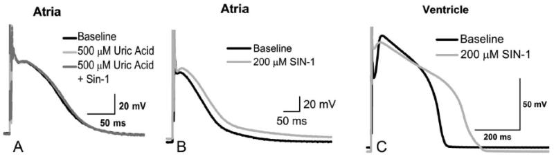
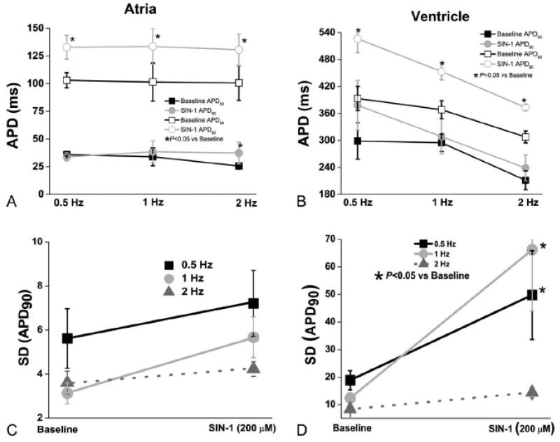
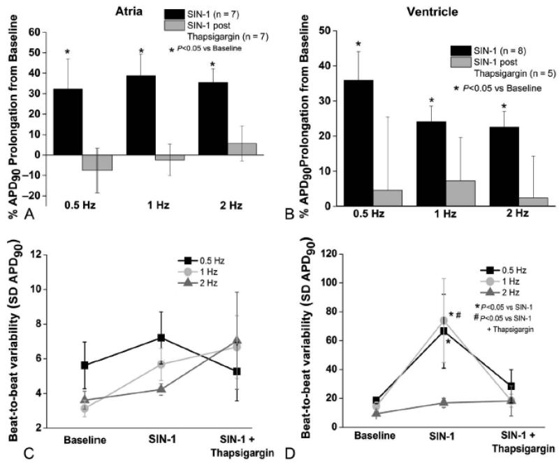
Oxidative stress has been implicated in the pathogenesis of heart failure and atrial fibrillation and can result in increased peroxynitrite production in the myocardium. Atrial and ventricular canine cardiac myocytes were superfused with 3-morpholinosydnonimine-N-ethylcarbamide (SIN-1), a peroxynitrite donor, to evaluate the acute electrophysiologic effects of peroxynitrite. Perforated whole-cell patch clamp techniques were used to record action potentials. SIN-1 (200 µM) increased the action potential duration (APD) in atrial and ventricular myocytes; however, in the atria, APD prolongation was rate independent, whereas in the ventricle APD, prolongation was rate dependent. In addition to prolongation of the action potential, beat-to-beat variability of repolarization was significantly increased in ventricular but not in atrial myocytes. We examined the contribution of intracellular calcium cycling to the effects of SIN-1 by treating myocytes with the SERCA blocker, thapsigargin (5-10 µM). Inhibition of calcium cycling prevented APD prolongation in the atrial and ventricular myocytes, and prevented the SIN-1-induced increase in ventricular beat-to-beat APD variability. Collectively, these data demonstrate that peroxynitrite affects atrial and ventricular electrophysiology differentially. A detailed understanding of oxidative modulation of electrophysiology in specific chambers is critical to optimize therapeutic approaches for cardiac diseases.
Figures
Similar articles
-
Calcium-activated potassium current modulates ventricular repolarization in chronic heart failure.PLoS One. 2014 Oct 1;9(10):e108824. doi: 10.1371/journal.pone.0108824. eCollection 2014. PLoS One. 2014. PMID: 25271970 Free PMC article.
-
Ranolazine improves abnormal repolarization and contraction in left ventricular myocytes of dogs with heart failure by inhibiting late sodium current.J Cardiovasc Electrophysiol. 2006 May;17 Suppl 1(Suppl 1):S169-S177. doi: 10.1111/j.1540-8167.2006.00401.x. J Cardiovasc Electrophysiol. 2006. PMID: 16686675 Free PMC article.
-
Peroxynitrite is a positive inotropic agent in atrial and ventricular fibres of the frog heart.J Physiol. 1999 Dec 1;521 Pt 2(Pt 2):375-88. doi: 10.1111/j.1469-7793.1999.00375.x. J Physiol. 1999. PMID: 10581309 Free PMC article.
-
Cyclic AMP and cyclic GMP independent stimulation of ventricular calcium current by peroxynitrite donors in guinea pig myocytes.J Cell Physiol. 2003 Nov;197(2):284-96. doi: 10.1002/jcp.10368. J Cell Physiol. 2003. PMID: 14502568
-
Remodeling of cardiac repolarization: mechanisms and implications of memory.Card Electrophysiol Rev. 2002 Sep;6(3):302-10. doi: 10.1023/a:1016349613464. Card Electrophysiol Rev. 2002. PMID: 12114856 Review.
Cited by
-
Calcium-activated potassium current modulates ventricular repolarization in chronic heart failure.PLoS One. 2014 Oct 1;9(10):e108824. doi: 10.1371/journal.pone.0108824. eCollection 2014. PLoS One. 2014. PMID: 25271970 Free PMC article.
-
Heart failure duration progressively modulates the arrhythmia substrate through structural and electrical remodeling.Life Sci. 2015 Feb 15;123:61-71. doi: 10.1016/j.lfs.2014.12.024. Epub 2015 Jan 14. Life Sci. 2015. PMID: 25596015 Free PMC article.
-
The NO/ONOO-cycle as the central cause of heart failure.Int J Mol Sci. 2013 Nov 13;14(11):22274-330. doi: 10.3390/ijms141122274. Int J Mol Sci. 2013. PMID: 24232452 Free PMC article. Review.
-
Cross-talk between lipid and protein carbonylation in a dynamic cardiomyocyte model of mild nitroxidative stress.Redox Biol. 2017 Apr;11:438-455. doi: 10.1016/j.redox.2016.12.028. Epub 2016 Dec 28. Redox Biol. 2017. PMID: 28086193 Free PMC article.
-
Nitric oxide: what's new to NO?Am J Physiol Cell Physiol. 2017 Mar 1;312(3):C254-C262. doi: 10.1152/ajpcell.00315.2016. Epub 2016 Dec 14. Am J Physiol Cell Physiol. 2017. PMID: 27974299 Free PMC article. Review.
References
-
- Han W, Fu S, Wei N, Xie B, Li W, Yang S, Li Y, Liang Z, Huo H. Nitric oxide overproduction derived from inducible nitric oxide synthase increases cardiomyocyte apoptosis in human atrial fibrillation. Int J Cardiol. 2008 Nov;130(2):165–173. - PubMed
-
- Sun Y. Oxidative stress and cardiac repair/remodeling following infarction. Am J Med Sci. 2007 Sep;334(3):197–205. - PubMed
-
- Kameda K, Matsunaga T, Abe N, Hanada H, Ishizaka H, Ono H, Saitoh M, Fukui K, Fukuda I, Osanai T, Okumura K. Correlation of oxidative stress with activity of matrix metalloproteinase in patients with coronary artery disease. Possible role for left ventricular remodelling. Eur Heart J. 2003 Dec;24(24):2180–2185. - PubMed
-
- Wattanapitayakul SK, Bauer JA. Oxidative pathways in cardiovascular disease: roles, mechanisms, and therapeutic implications. Pharmacol Ther. 2001 Feb;89(2):187–206. - PubMed
Publication types
MeSH terms
Substances
Grants and funding
LinkOut - more resources
Full Text Sources
Other Literature Sources
