

Poly(β-amino ester) nanoparticle delivery of TP53 has activity against small cell lung cancer in vitro and in vivo
- PMID: 23364678
- PMCID: PMC3624031
- DOI: 10.1158/1535-7163.MCT-12-0956
Poly(β-amino ester) nanoparticle delivery of TP53 has activity against small cell lung cancer in vitro and in vivo
Abstract
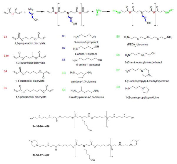
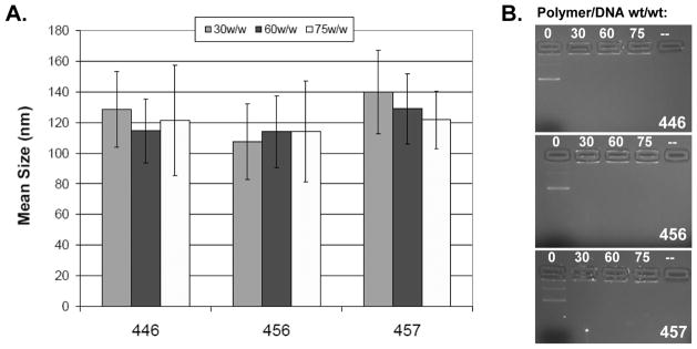
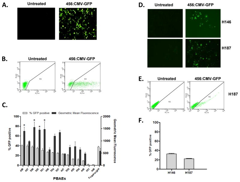
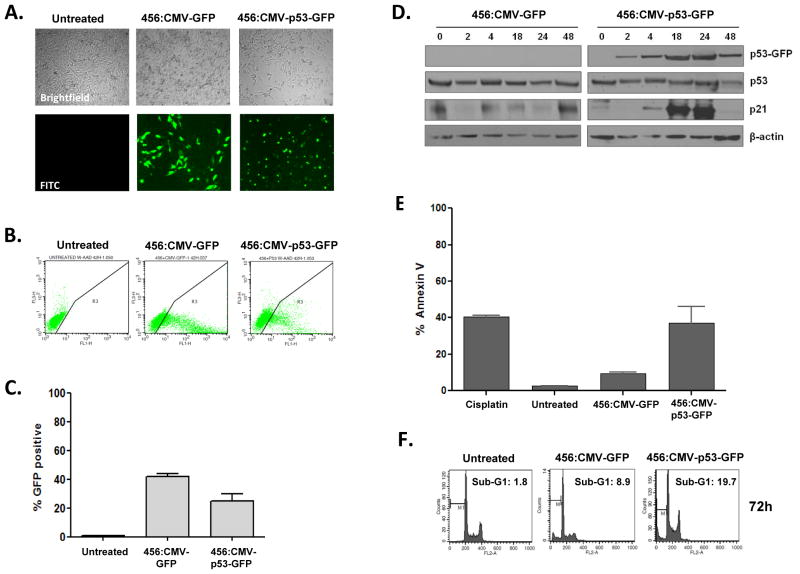
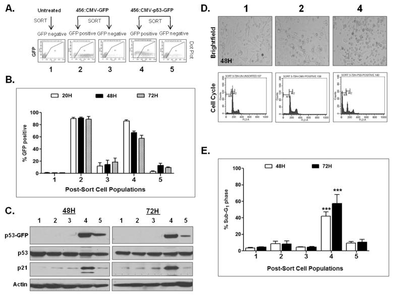
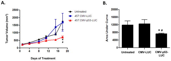
Small cell lung cancer (SCLC) is an aggressive disease with one of the highest case-fatality rates among cancer. The recommended therapy for SCLCs has not changed significantly over the past 30 years; new therapeutic approaches are a critical need. TP53 is mutated in the majority of SCLC cases and its loss is required in transgenic mouse models of the disease. We synthesized an array of biodegradable poly(β-amino ester) (PBAE) polymers that self-assemble with DNA and assayed for transfection efficiency in the p53-mutant H446 SCLC cell line using high-throughput methodologies. Two of the top candidates were selected for further characterization and TP53 delivery in vitro and in vivo. Nanoparticle delivery of TP53 resulted in expression of exogenous p53, induction of p21, induction of apoptosis, and accumulation of cells in sub-G1 consistent with functional p53 activity. Intratumoral injection of subcutaneous H446 xenografts with polymers carrying TP53 caused marked tumor growth inhibition. This is the first demonstration of TP53 gene therapy in SCLC using nonviral polymeric nanoparticles. This technology may have general applicability as a novel anticancer strategy based on restoration of tumor suppressor gene function.
Conflict of interest statement
Figures
Similar articles
-
Synthesis and application of poly(ethylene glycol)-co-poly(β-amino ester) copolymers for small cell lung cancer gene therapy.Acta Biomater. 2016 Sep 1;41:293-301. doi: 10.1016/j.actbio.2016.05.040. Epub 2016 Jun 1. Acta Biomater. 2016. PMID: 27262740 Free PMC article.
-
Enhanced tumor suppression by adenoviral PTEN gene therapy combined with cisplatin chemotherapy in small-cell lung cancer.Cancer Gene Ther. 2013 Apr;20(4):251-9. doi: 10.1038/cgt.2013.14. Epub 2013 Mar 8. Cancer Gene Ther. 2013. PMID: 23470565
-
MicroRNA-886-3P functions as a tumor suppressor in small cell lung cancer.Cancer Biol Ther. 2018;19(12):1185-1192. doi: 10.1080/15384047.2018.1491505. Epub 2018 Sep 19. Cancer Biol Ther. 2018. PMID: 30230945 Free PMC article.
-
Glucocorticoid receptor over-expression promotes human small cell lung cancer apoptosis in vivo and thereby slows tumor growth.Endocr Relat Cancer. 2010 Feb 18;17(1):203-13. doi: 10.1677/ERC-09-0241. Print 2010 Mar. Endocr Relat Cancer. 2010. PMID: 20015838 Free PMC article.
-
Advances in Small-Cell Lung Cancer (SCLC) Translational Research.Cold Spring Harb Perspect Med. 2021 Apr 1;11(4):a038240. doi: 10.1101/cshperspect.a038240. Cold Spring Harb Perspect Med. 2021. PMID: 32513672 Free PMC article. Review.
Cited by
-
Synthesis and application of poly(ethylene glycol)-co-poly(β-amino ester) copolymers for small cell lung cancer gene therapy.Acta Biomater. 2016 Sep 1;41:293-301. doi: 10.1016/j.actbio.2016.05.040. Epub 2016 Jun 1. Acta Biomater. 2016. PMID: 27262740 Free PMC article.
-
Polymeric nanoparticle-based delivery of TRAIL DNA for cancer-specific killing.Bioeng Transl Med. 2016 Jun;1(2):149-159. doi: 10.1002/btm2.10019. Bioeng Transl Med. 2016. PMID: 28349127 Free PMC article.
-
In vivo gene delivery mediated by non-viral vectors for cancer therapy.J Control Release. 2020 Sep 10;325:249-275. doi: 10.1016/j.jconrel.2020.06.038. Epub 2020 Jul 4. J Control Release. 2020. PMID: 32634464 Free PMC article. Review.
-
Non-viral nucleic acid containing nanoparticles as cancer therapeutics.Expert Opin Drug Deliv. 2016 Oct;13(10):1475-87. doi: 10.1080/17425247.2016.1190707. Epub 2016 Jun 6. Expert Opin Drug Deliv. 2016. PMID: 27248202 Free PMC article. Review.
-
Biodegradable polymeric nanoparticles show high efficacy and specificity at DNA delivery to human glioblastoma in vitro and in vivo.ACS Nano. 2014 May 27;8(5):5141-53. doi: 10.1021/nn501197v. Epub 2014 Apr 29. ACS Nano. 2014. PMID: 24766032 Free PMC article.
References
Publication types
MeSH terms
Substances
Grants and funding
LinkOut - more resources
Full Text Sources
Other Literature Sources
Medical
Research Materials
Miscellaneous
