. 2013 Jan 30;11(2):332-49.
doi: 10.3390/md11020332.

Iodinin (1,6-dihydroxyphenazine 5,10-dioxide) from Streptosporangium sp. induces apoptosis selectively in myeloid leukemia cell lines and patient cells

Affiliations

Iodinin (1,6-dihydroxyphenazine 5,10-dioxide) from Streptosporangium sp. induces apoptosis selectively in myeloid leukemia cell lines and patient cells

Lene E Myhren et al. Mar Drugs. .

Abstract

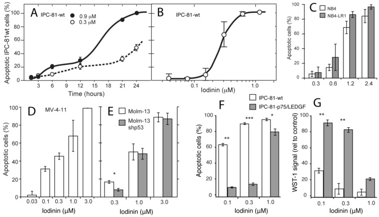
Despite recent improvement in therapy, acute myeloid leukemia (AML) is still associated with high lethality. In the presented study, we analyzed the bioactive compound iodinin (1,6-dihydroxyphenazine 5,10-dioxide) from a marine actinomycetes bacterium for the ability to induce cell death in a range of cell types. Iodinin showed selective toxicity to AML and acute promyelocytic (APL) leukemia cells, with EC50 values for cell death up to 40 times lower for leukemia cells when compared with normal cells. Iodinin also successfully induced cell death in patient-derived leukemia cells or cell lines with features associated with poor prognostic such as FLT3 internal tandem duplications or mutated/deficient p53. The cell death had typical apoptotic morphology, and activation of apoptotic signaling proteins like caspase-3. Molecular modeling suggested that iodinin could intercalate between bases in the DNA in a way similar to the anti-cancer drug daunorubicin (DNR), causing DNA-strand breaks. Iodinin induced apoptosis in several therapy-resistant AML-patient blasts, but to a low degree in peripheral blood leukocytes, and in contrast to DNR, not in rat cardiomyoblasts. The low activity towards normal cell types that are usually affected by anti-leukemia therapy suggests that iodinin and related compounds represent promising structures in the development of anti-cancer therapy.


Figures

Figure 1
Figure 1
Iodinin is a potent cell death inducer in acute promyelocytic leukemia (APL) and acute myeloid leukemia (AML) cell lines. (A) Rat acute promyelocytic leukemia cells (IPC-81) were incubated with iodinin for various periods of time, fixed in buffered formaldehyde with the DNA-dye Hoechst 33342, and cell death was scored by differential interference contrast and UV-microscopy. (BE) APL and AML cells were treated with increasing concentrations of iodinin for 24 h fixed in buffered formaldehyde and cell death was scored as described above. (F and G) IPC-81-wt cells or expressing p75/LEDGF were given daily pulses of anthracycline daunorubicin (DNR) for three days as described in the Experimental Section. The metabolic activity was measured by the WST-1 assay, and apoptosis as described for panel (A). Untreated cells or cells treated with solvent had always less than 4% apoptosis. The data are average and SEM of 3–5 experiments. Asterisks denote significance at p < 0.05 (*), <0.01 (**), <0.005 (***), t-test, for comparison of different strains of the same cell line (C, E, F and G).
Figure 2
Figure 2
Iodinin induces apoptotic cell death in leukemia cells. (AD) Transmission electron micrographs of IPC-81 cells treated with solvent (DMSO, A), or 0.3 μM of iodinin (BD) for 21 h. N is nucleus, M is mitochondria, and FC is fragmented and condensed DNA. (E) Modulations of proteins in iodinin- and DNR-mediated leukemia cell death. NB4 cells were treated with the given concentrations of DNR or iodinin for 6 h. Cell extracts were immuno-blotted and probed for caspase 3, γH2AX, and β-actin, as described in the Experimental Section.
Figure 3
Figure 3
Structural similarities between iodinin and DNR. Pharmacophore model of iodinin (A), common pharmacophore features of iodinin and DNR (B), and suggested intercalation of iodinin (colored by atom type) with DNA (green and grey) (C). DNR is also shown in orange, overlapping with iodinin. The pharmacophore models were created with the Phase module of the Schrödinger™ software, whereas the suggested interaction of iodinin with DNA was prepared in Discovery Studio, based on the structural alignment (B) and a previously published interaction of DNR with DNA [27].
Figure 4
Figure 4
Iodinin induces apoptosis in leukemia patient blasts. Blasts isolated from peripheral blood samples from six leukemia patients were treated with iodinin (0.3, 1 or 3 μM) or DNR (0.2 or 0.5 μM) for 18 h. Samples were assessed for drug-induced cell death by FACS analysis of AnnexinV and PI labeling. The background (control) was subtracted from the data. See Table 2 for description of the patient samples.
Figure 5
Figure 5
Iodinin exhibit low toxicity towards normal cells and tissues. (A) Rat cardiomyoblasts (H9C2) were incubated with iodinin or DNR for 24 h before viability was assessed by microscopic evaluation of surface morphology. (B) Peripheral blood leukocytes were incubated with the indicated concentrations of iodinin or DNR for 24 h before FACS analysis of AnnexinV and PI staining. (C) Blood platelets were incubated (15 min) with increasing concentrations of the thrombin receptor agonist peptide (TRAP) with or without iodinin or DNR and activation was determined as described in the Experimental section. Platelets incubated with 20 μM TRAP was set as 100% activation. The data are average of three to five experiments and SEM (A) and (C), or two experiments and each measurement (B). Asterisks denote significance at p < 0.05 (*), <0.01 (**), <0.005 (***), t-test in (AC). (D and E) Histological appearance of duodenum of mice given vehicle, (D) or iodinin (10mg/kg, E) daily for three days. Organs were collected the day after the last treatment. See Experimental Section for further details.
Figure S1
Figure S1
UV-DAD spectra of purified iodinin.
Figure S2
Figure S2
UV profile of purified iodinin produced by isolate MP55-27. UV absorption (nm) at UV max and % of UV max stated in the figure.
Figure S3
Figure S3
LC-TOF (A) and LC-Trap spectra (B) of iodinin. Two main ions were detected, and the accurate mass of the most abundant isotope peak in each spectrum is stated above each MS-spectrum. The two main ions represent iodinin monomer and the Na-adduct of the iodinin dimer. The spectrum inserted in (B) is the MS/MS spectrum of the Na-adduct of the iodinin dimer (m/z = 509).

Similar articles

Cited by

References

    1. Fenaux P., Chastang C., Chevret S., Sanz M., Dombret H., Archimbaud E., Fey M., Rayon C., Huguet F., Sotto J.J., et al. A randomized comparison of all transretinoic acid (ATRA) followed by chemotherapy and ATRA plus chemotherapy and the role of maintenance therapy in newly diagnosed acute promyelocytic leukemia. Blood. 1999;94:1192–1200. - PubMed
    1. Mayer R.J., Davis R.B., Schiffer C.A., Berg D.T., Powell B.L., Schulman P., Omura G.A., Moore J.O., McIntyre O.R., Frei E., III. Intensive postremission chemotherapy in adults with acute myeloid leukemia. N. Engl. J. Med. 1994;331:896–903. doi: 10.1056/NEJM199410063311402. - DOI - PubMed
    1. Mehta A., Hoffbrand V. Haematology at a Glance. 3rd. Wiley-Blackwell; Oxford, UK: 2010.
    1. Burnett A., Wetzler M., Lowenberg B. Therapeutic advances in acute myeloid leukemia. J. Clin. Oncol. 2011;29:487–494. doi: 10.1200/JCO.2010.30.1820. - DOI - PubMed
    1. Jemal A., Clegg L.X., Ward E., Ries L.A., Wu X., Jamison P.M., Wingo P.A., Howe H.L., Anderson R.N., Edwards B.K. Annual report to the nation on the status of cancer, 1975-2001, with a special feature regarding survival. Cancer. 2004;101:3–27. doi: 10.1002/cncr.20288. - DOI - PubMed

MeSH terms