. 2013 Mar 22;288(12):8074-8084.
doi: 10.1074/jbc.M112.432773. Epub 2013 Jan 30.

Nuclear calcium signaling regulates nuclear export of a subset of class IIa histone deacetylases following synaptic activity

Affiliations

Nuclear calcium signaling regulates nuclear export of a subset of class IIa histone deacetylases following synaptic activity

Friederike Schlumm et al. J Biol Chem. .

Abstract

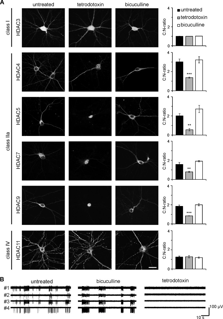
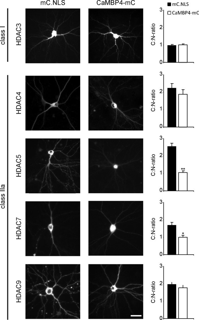
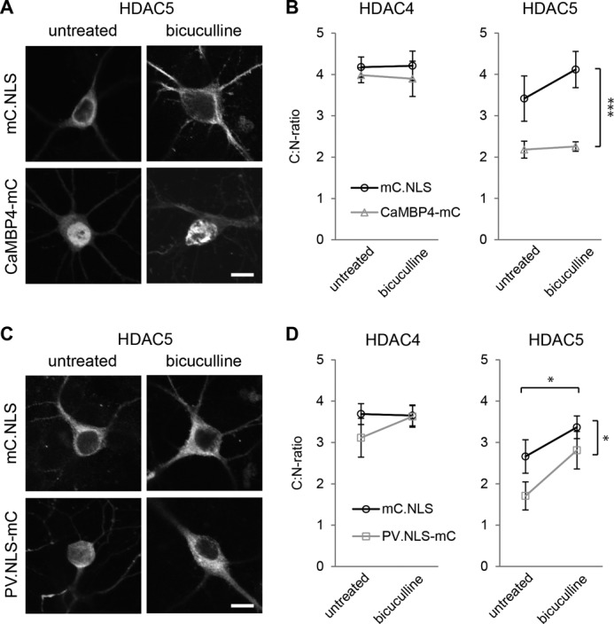
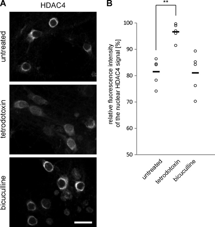
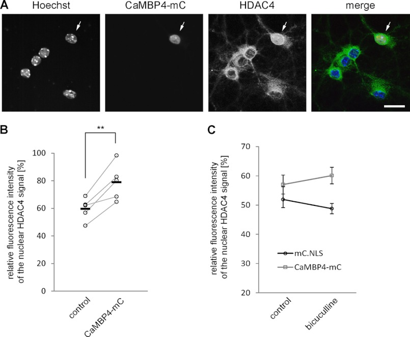
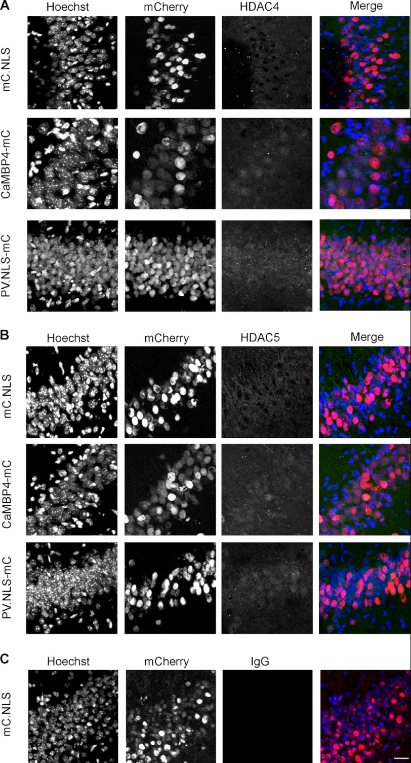
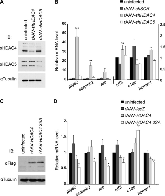
In neurons, dynamic changes in the subcellular localization of histone deacetylases (HDACs) are thought to contribute to signal-regulated gene expression. Here we show that in mouse hippocampal neurons, synaptic activity-dependent nucleo-cytoplasmic shuttling is a common feature of all members of class IIa HDACs, which distinguishes them from other classes of HDACs. Nuclear calcium, a key regulator in neuronal gene expression, is required for the nuclear export of a subset of class IIa HDACs. We found that inhibition of nuclear calcium signaling using CaMBP4 or increasing the nuclear calcium buffering capacity by means of expression of a nuclear targeted version of parvalbumin (PV.NLS-mC) led to a build-up of HDAC4 and HDAC5 in the cell nucleus, which in the case of PV.NLS-mC can be reversed by nuclear calcium transients triggered by bursts of action potential firing. A similar nuclear accumulation of HDAC4 and HDAC5 was observed in vivo in the mouse hippocampus following stereotaxic delivery of recombinant adeno-associated viruses expressing either CaMBP4 or PV.NLS-mC. The modulation of HDAC4 activity either by RNA interference-mediated reduction of HDAC4 protein levels or by expression of a constitutively nuclear localized mutant of HDAC4 leads to changes in the mRNA levels of several nuclear calcium-regulated genes with known functions in acquired neuroprotection (atf3, serpinb2), memory consolidation (homer1, arc), and the development of chronic pain (ptgs2, c1qc). These results identify nuclear calcium as a regulator of nuclear export of HDAC4 and HDAC5. The reduction of nuclear localized HDACs represents a novel transcription-promoting pathway stimulated by nuclear calcium.


Figures

FIGURE 1.
FIGURE 1.
Nucleo-cytoplasmic shuttling of class IIa HDACs is dependent on neuronal activity. A, representative micrographs of cultured hippocampal neurons transfected with epitope-tagged HDAC constructs (HDAC3-HA, HDAC4-FLAG, HDAC5-FLAG, HDAC7-HA, HDAC9-HA, and HDAC11-HA) and either left untreated or treated with TTX or bicuculline as indicated. Scale bar is 20 μm. Graphs show the ratio between cytoplasmic and nuclear localization. 400–1000 cells were analyzed for each tested HDAC and experimental condition from a minimum of three independent preparations. Statistically significant differences are indicated with asterisks (***, p < 0.001; **, p < 0.01, one-way ANOVA, Dunnett's post hoc test). Error bars, S.E. B, typical examples of MEA recordings obtained from untreated cultured hippocampal neurons (left), and neurons after exposure to bicuculline (50 μm, center), or TTX (1 μm, right). Simultaneous recordings from four separate electrodes are shown in each panel.
FIGURE 2.
FIGURE 2.
Nuclear calcium differentially regulates the shuttling of class IIa HDACs. Representative images show hippocampal neurons transfected with HDAC constructs with expression vector for CaMBP4-mCherry (CaMBP4-mC) or mCherry-NLS (mC.NLS) as indicated. HDACs subcellular localization was analyzed only in CaMBP4- or mC.NLS-expressing cells, selected by mCherry expression. Scale bar is 20 μm. Graphs show the ratio between cytoplasmic and nuclear localization. 140–700 cells were analyzed for each condition from a minimum of three independent experiments. Statistically significant differences are indicated with asterisks (**, p < 0.01; *, p < 0.05, two-sided Student's t test). Error bars, S.E.
FIGURE 3.
FIGURE 3.
Increasing neuronal activity can overcome parvalbumin-dependent but not CaMBP4-dependent block of HDAC5 nuclear export. A, representative images of hippocampal neurons transfected with HDAC5 and with mCherry (mC.NLS) or CaMBP4-mCherry (CaMBP4-mC) as indicated. Neurons were either left untreated or treated with bicuculline. Scale bar is 10 μm. B, quantitative analysis of the subcellular localization of HDAC4 or HDAC5 in hippocampal neurons. Neurons were transfected with HDAC4 or HDAC5 and with mCherry or CaMBP4 and treated with bicuculline or not as indicated. Graphs show the ratio between cytoplasmic and nuclear localization. In total, 140–400 cells were analyzed for each condition from a minimum of five independent experiments. C, representative images of hippocampal neurons transfected with HDAC5 and with mCherry (mC.NLS) or parvalbumin-NLS-mCherry (PV.NLS-mC) as indicated. Neurons were either left untreated or treated with bicuculline. Scale bar is 10 μm. D, quantitative analysis of the subcellular localization of HDAC4 or HDAC5 in hippocampal neurons. Neurons were transfected with HDAC4 or HDAC5 and with mCherry or PV.NLS-mC and treated with bicuculline or not as indicated. Graphs show the ratio between cytoplasmic and nuclear localization. In total, 140–200 cells were analyzed for each condition from a minimum of five independent experiments. Statistically significant differences are indicated with asterisks (***, p < 0.001; *, p < 0.05, two-way ANOVA). Error bars, S.E.
FIGURE 4.
FIGURE 4.
Nucleo-cytoplasmic shuttling of endogenous HDAC4 is dependent on neuronal activity. A, representative images of hippocampal neurons left untreated or treated with tetrodotoxin or bicuculline and immunostained for endogenous HDAC4. Scale bar is 20 μm. B, quantitative measurements of the relative fluorescent intensity of HDAC4 signal in the nucleus compared with the signal of the cytosol. Each circle represents an independent experiment. Horizontal bars show mean values (control, 81.5 ± 2.3%; bicuculline, 81.0 ± 3.4%, p = 0.99; TTX, 96.6 ± 1.4% p = 0.002; one-way ANOVA, Dunnett's post hoc test, F = 12.34). Statistically significant difference is indicated with asterisks (**).
FIGURE 5.
FIGURE 5.
Shuttling of endogenous HDAC4 is responsive to nuclear calcium signaling. A, representative images showing the subcellular localization of endogenous HDAC4 in hippocampal neuron transfected with CaMBP4-mCherry (arrow) and nontransfected surrounding control cells. Scale bar is 20 μm. B, quantitative measurements of the relative fluorescent intensity of HDAC4 signal in the nucleus compared with the signal of the cytosol in untransfected (control) and CaMBP4-expressing cells. Each circle represents an independent experiment. Horizontal bars show mean values (control, 59.6 ± 3.6%; CaMBP4, 78.9 ± 5.9% p = 0.007 paired Student's t test). Statistically significant difference is indicated with asterisks (**). C, quantitative measurements of the relative fluorescent intensity of HDAC4 signal in the nucleus compared with the signal of the cytosol in mC.NLS- and CaMBP4-mC-expressing cells. Neurons were left untreated or treated with bicuculline and immunostained for endogenous HDAC4. The averaged results of three independent experiments are shown. Error bars, S.E.
FIGURE 6.
FIGURE 6.
Nuclear calcium controls HDAC4 and -5 nucleo-cytoplasmic shuttling in vivo. A, immunohistochemistry of HDAC4 in the CA1 region of the hippocampus of adult mice stereotaxically injected with rAAV-mCherry-NLS (mC.NLS), rAAV-CAMBP4-mCherry (CaMBP4-mC) or with rAAV-parvalbumin.NLS-mCherry (PV.NLS-mC) using an anti-HDAC4 antibody, Hoechst to visualize the nuclei, and mCherry fluorescence to detect virus expression. B, immunohistochemistry of HDAC5 in the CA1 region of the hippocampus of adult mice stereotaxically injected with rAAV-mCherry-NLS (mC.NLS), rAAV-CAMBP4-mCherry (CaMBP4-mC), or with rAAV-parvalbumin.NLS-mCherry (PV.NLS-mC) using an anti-HDAC5 antibody, Hoechst to visualize the nuclei, and mCherry fluorescence to detect virus expression. C, immunohistochemistry of the CA1 region of the hippocampus of adult mice stereotaxically injected with rAAV-mCherry.NLS (mC.NLS), using a normal rabbit IgG antibody to test the specificity of the immunostainings shown in A and B, Hoechst to visualize the nuclei, and mCherry fluorescence to detect virus expression. Scale bar is 20 μm.
FIGURE 7.
FIGURE 7.
HDAC4 regulates expression of genes involved in neuroadaptations. A, immunoblot analysis of uninfected hippocampal neurons and of hippocampal neurons infected with the indicated rAAVs. Tubulin immunoblot is shown as control for protein loading. B, QRT-PCR analysis of expression of ptgs2, serpinb2, arc, atf3, c1qc, and homer1 in uninfected hippocampal neurons and in hippocampal neurons infected with rAAV-shSCR, rAAV-shHDAC4, or with rAAV-shHDAC5 (n = 5). ptgs2, arc, and serpinb2 values refer to the axis on the left. C, immunoblot analysis of uninfected hippocampal neurons and of hippocampal neurons infected with the indicated rAAVs. rAAV-HDAC4 and rAAV-HDAC4 3SA all carry a FLAG cassette which was used for detection via an anti-FLAG antibody. Tubulin immunoblot is shown as control for protein loading. D, QRT-PCR analysis of ptgs2, serpinb2, arc, atf3, c1qc, and homer1 expression in uninfected hippocampal neurons and in hippocampal neurons infected with rAAVs giving rise to the indicated proteins (n = 5). Statistically significant differences are indicated with asterisks (***, p < 0.0005; **, p < 0.05; *, p < 0.05 one-way ANOVA, Dunnett's post hoc test). Error bars, S.E.

Similar articles

Cited by

References

    1. Greer P. L., Greenberg M. E. (2008) From synapse to nucleus: calcium-dependent gene transcription in the control of synapse development and function. Neuron 59, 846–860 - PubMed
    1. Hagenston A. M., Bading H. (2011) Calcium signaling in synapse-to-nucleus communication. Cold Spring Harb. Perspect. Biol. 3, a004564. - PMC - PubMed
    1. Hardingham G. E., Chawla S., Johnson C. M., Bading H. (1997) Distinct functions of nuclear and cytoplasmic calcium in the control of gene expression. Nature 385, 260–265 - PubMed
    1. Limbäck-Stokin K., Korzus E., Nagaoka-Yasuda R., Mayford M. (2004) Nuclear calcium/calmodulin regulates memory consolidation. J. Neurosci. 24, 10858–10867 - PMC - PubMed
    1. Papadia S., Stevenson P., Hardingham N. R., Bading H., Hardingham G. E. (2005) Nuclear Ca2+ and the cAMP response element-binding protein family mediate a late phase of activity-dependent neuroprotection. J. Neurosci. 25, 4279–4287 - PMC - PubMed

Publication types

MeSH terms