. 2013 Mar 22;288(12):8627-8635.
doi: 10.1074/jbc.M112.441949. Epub 2013 Jan 30.

Secreted progranulin is a homodimer and is not a component of high density lipoproteins (HDL)

Affiliations

Secreted progranulin is a homodimer and is not a component of high density lipoproteins (HDL)

Andrew D Nguyen et al. J Biol Chem. .

Abstract

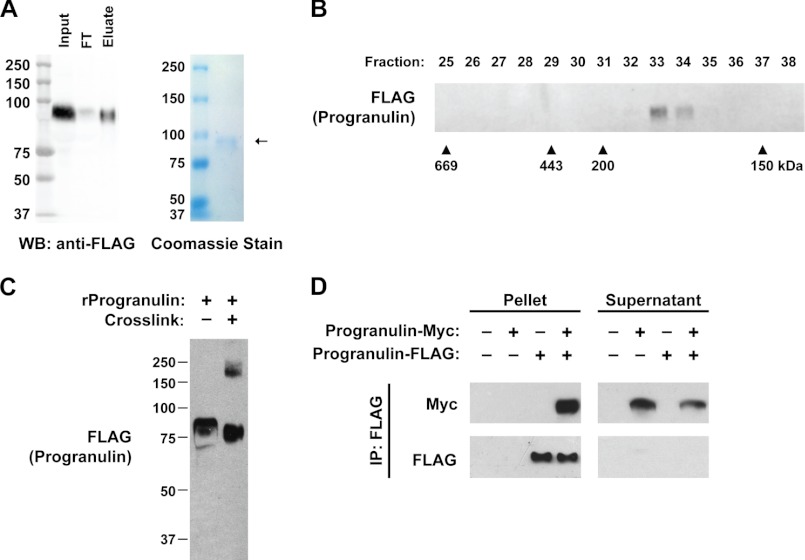
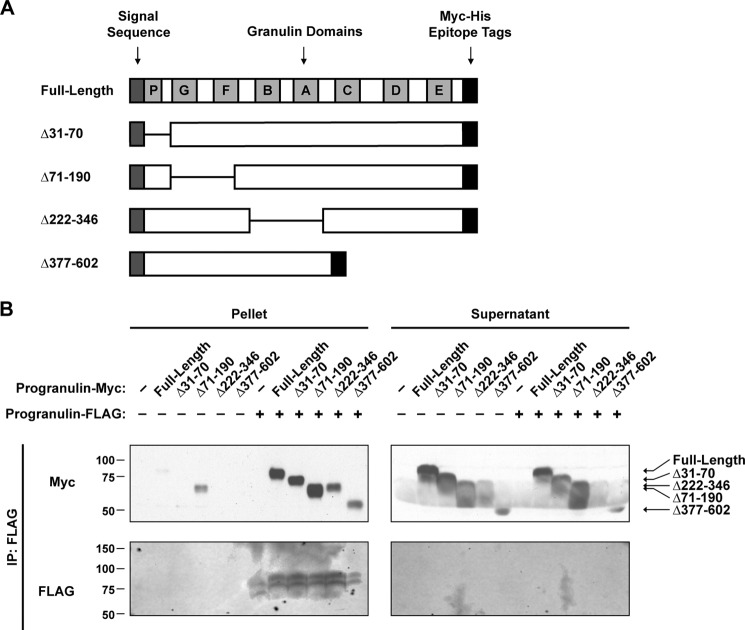
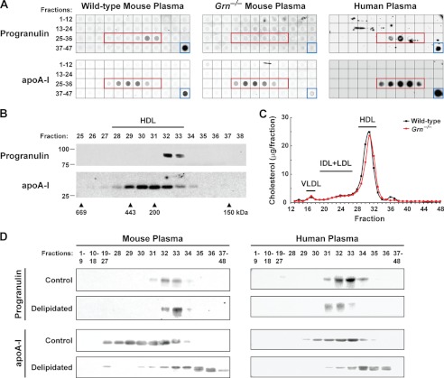
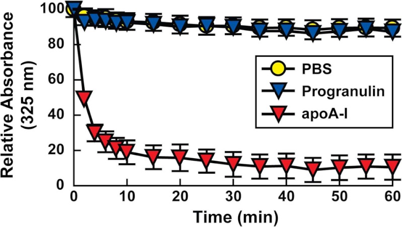
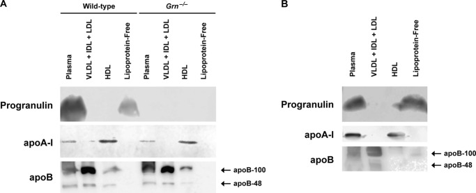
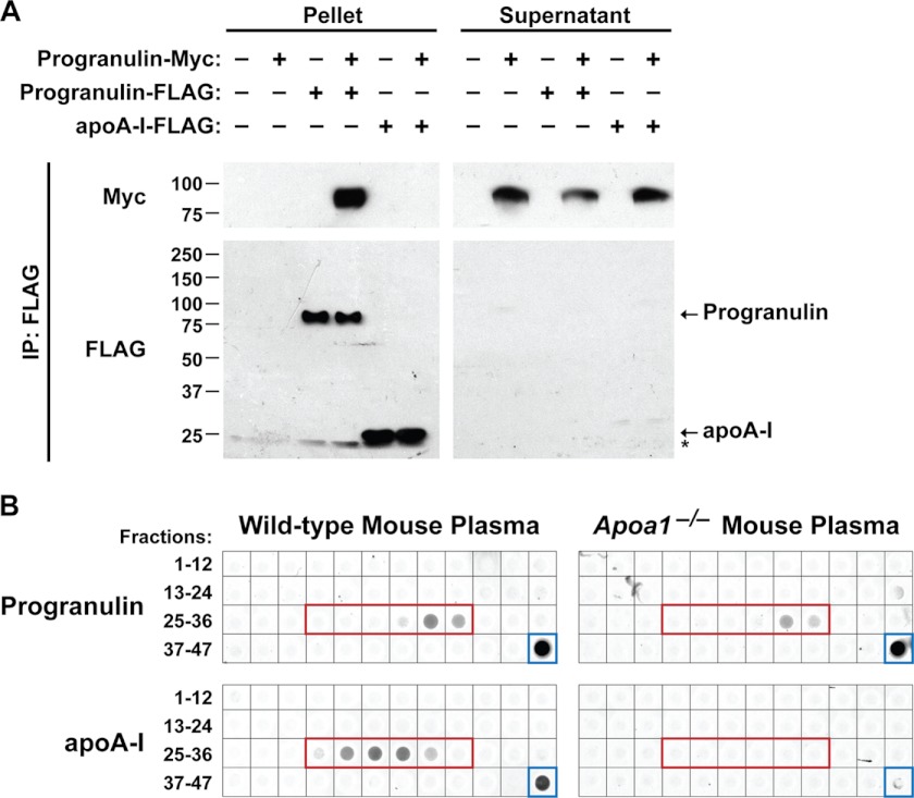
Progranulin is a secreted glycoprotein, and the GRN gene is mutated in some cases of frontotemporal dementia. Progranulin has also been implicated in cell growth, wound healing, inflammation, and cancer. We investigated the molecular nature of secreted progranulin and provide evidence that progranulin exists as a homodimer. Although recombinant progranulin has a molecular mass of ∼85 kDa by SDS-PAGE, it elutes in fractions corresponding to ∼170-180 kDa by gel-filtration chromatography. Additionally, recombinant progranulin can be intermolecularly cross-linked, yielding a complex corresponding to a dimer (∼180 kDa), and progranulins containing different epitope tags physically interact. In plasma, progranulin similarly forms complexes of ∼180-190 kDa. Although progranulin partially co-fractionated with high density lipoproteins (HDL) by gel-filtration chromatography, we found no evidence that progranulin in mouse or human plasma is a component of HDL either by ultracentrifugation or by lipid binding assays. We conclude that circulating progranulin exists as a dimer and is not likely a component of HDL.


Figures

FIGURE 1.
FIGURE 1.
Recombinant progranulin is a homodimer. A, purification of secreted recombinant human progranulin from conditioned media of transfected HEK293T cells is shown. Anti-FLAG Western blot (WB, left) and Coomassie-stained gel (right) of purified progranulin are shown. FT, flow-through. B, recombinant human progranulin migrates at ∼170–180 kDa. Western blot of gel-filtration fractions. Similar results were obtained in two independent experiments. C, cross-linking partially shifts the size of recombinant progranulin from ∼85 to ∼180 kDa. Shown is a Western blot of recombinant human progranulin subjected to cross-linking with disuccinimidyl suberate. D, expressed progranulin-FLAG physically interacts with progranulin-Myc. Co-immunoprecipitation (IP) of conditioned medium from transfected HEK293T cells with anti-FLAG magnetic beads is shown. These results are from a representative experiment that was performed three times with similar results.
FIGURE 2.
FIGURE 2.
Mapping of the dimerization domain of progranulin. A, a schematic representation of deletion mutants of mouse progranulin is shown. B, mapping of dimerization domain of progranulin in co-immunoprecipitation (IP) assays is shown. Conditioned media from transfected HEK293T cells was subjected to immunoprecipitation with anti-FLAG magnetic beads. Similar results were obtained in three independent experiments.
FIGURE 3.
FIGURE 3.
Circulating progranulin is found in complexes of ∼180–190 kDa, partially co-elutes with HDL by gel-filtration, and is not associated with lipids. A, progranulin in mouse and human plasma is in a complex of ∼180–190 kDa and partially co-elutes with HDL. Dot blots of gel-filtration fractions 1–47 and plasma (blue box) from wild-type mice (left panel), Grn−/− mice (middle panel), and a human subject (right panel) are shown. The fractions corresponding to HDL are indicated by the red box. B, progranulin in mouse plasma partially co-elutes with HDL. Western blots of gel-filtration fractions 25–38 from wild-type mouse plasma are shown. These results are from a representative experiment that was performed four times with similar results. C, cholesterol profiles of plasma from wild-type (black line) and Grn−/− (red line) mice are shown. IDL, intermediate density lipoproteins. D, circulating progranulin is not associated with lipids. Delipidation of mouse (left panel) and human (right panel) plasma results in a shift in the gel-filtration elution profile of apoA-I but not in that of progranulin. Western blots of gel-filtration fractions 1–48 for control and delipidated mouse plasma. Similar results were obtained in two independent experiments.
FIGURE 4.
FIGURE 4.
Recombinant progranulin does not reorganize DMPC vesicles. A phospholipid clearance assay using recombinant human progranulin and purified human apoA-I is shown. ApoA-I cleared the DMPC vesicles (p < 0.001 compared with PBS), but progranulin did not (p > 0.05, compared with PBS). Data are presented as the mean ± S.D. Comparisons between groups were made by one-way analysis of variance followed by the Tukey-Kramer post-hoc test. Similar results were obtained in three independent experiments.
FIGURE 5.
FIGURE 5.
Circulating progranulin is not a component of murine or human lipoproteins isolated by ultracentrifugation. Western blots of plasma and lipoprotein fractions isolated from mouse (A) and human (B) plasma are shown. These results are from representative experiments that were performed three times (A) and two times (B) with similar results. IDL, intermediate density lipoproteins.
FIGURE 6.
FIGURE 6.
Progranulin and apoA-I do not physically interact. A, progranulin and apoA-I do not physically interact in co-immunoprecipitation (IP) assays. Conditioned medium from transfected HEK293T cells was subjected to immunoprecipitation with anti-FLAG magnetic beads. The asterisk indicates a cross-reactive band. These results are from a representative experiment that was performed six times with similar results. B, gel-filtration elution profile of progranulin is not affected by loss of apoA-I. Dot blots of gel-filtration fractions 1–47 and plasma (blue box) from wild-type (left) and Apoa1−/− (right) mice are shown. The fractions corresponding to HDL are indicated by the red box. Similar results were obtained in two independent experiments.

References

    1. He Z., Ong C. H., Halper J., Bateman A. (2003) Progranulin is a mediator of the wound response. Nat. Med. 9, 225–229 - PubMed
    1. Cenik B., Sephton C. F., Cenik B. K., Herz J., Yu G. (2012) Progranulin. A proteolytically processed protein at the crossroads of inflammation and neurodegeneration. J. Biol. Chem. 287, 32298–32306 - PMC - PubMed
    1. Ryan C. L., Baranowski D. C., Chitramuthu B. P., Malik S., Li Z., Cao M., Minotti S., Durham H. D., Kay D. G., Shaw C. A., Bennett H. P., Bateman A. (2009) Progranulin is expressed within motor neurons and promotes neuronal cell survival. BMC Neuroscience 10, 130. - PMC - PubMed
    1. He Z., Bateman A. (1999) Progranulin gene expression regulates epithelial cell growth and promotes tumor growth in vivo. Cancer Res. 59, 3222–3229 - PubMed
    1. Ong C. H., He Z., Kriazhev L., Shan X., Palfree R. G., Bateman A. (2006) Regulation of progranulin expression in myeloid cells. Am. J. Physiol. Regul. Integr. Comp. Physiol. 291, R1602–E1612 - PubMed

Publication types

MeSH terms