. 2013 Mar 22;288(12):8101-8110.
doi: 10.1074/jbc.M112.431148. Epub 2013 Jan 30.

Nuclease activity of the human SAMHD1 protein implicated in the Aicardi-Goutieres syndrome and HIV-1 restriction

Affiliations

Nuclease activity of the human SAMHD1 protein implicated in the Aicardi-Goutieres syndrome and HIV-1 restriction

Natalia Beloglazova et al. J Biol Chem. .

Abstract

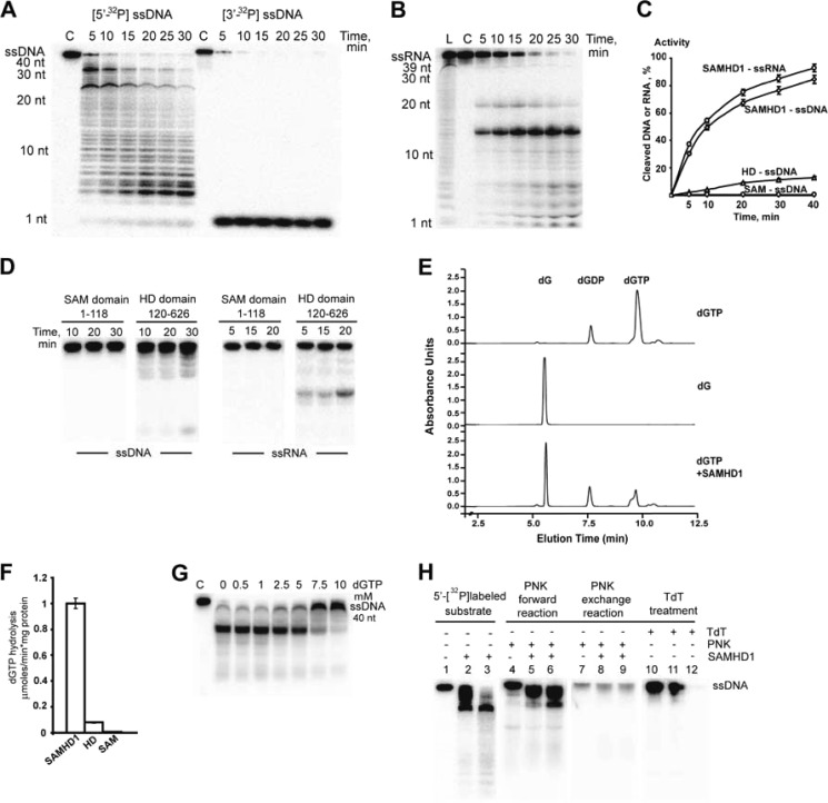
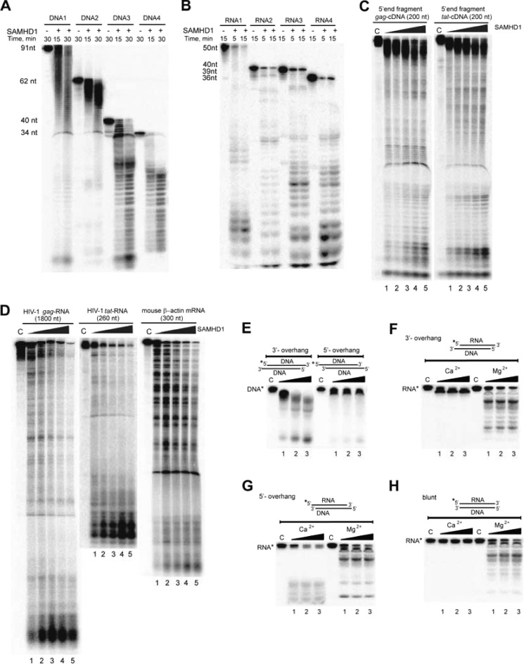
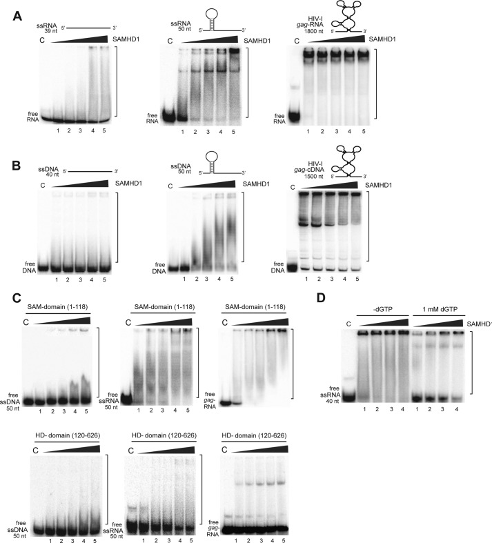
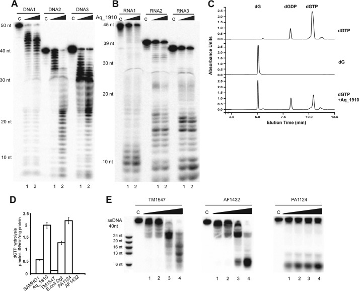
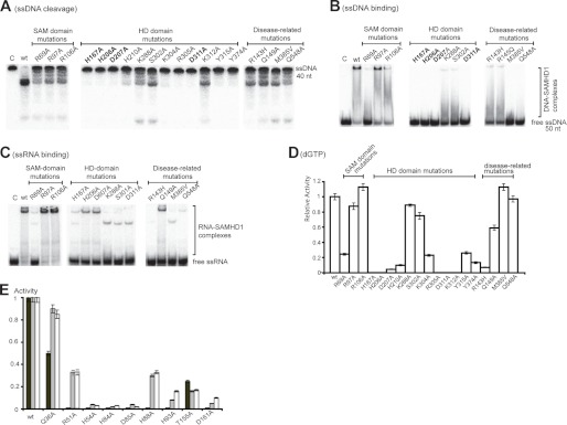
The human HD domain protein SAMHD1 is implicated in the Aicardi-Goutières autoimmune syndrome and in the restriction of HIV-1 replication in myeloid cells. Recently, this protein has been shown to possess dNTP triphosphatase activity, which is proposed to inhibit HIV-1 replication and the autoimmune response by hydrolyzing cellular dNTPs. Here, we show that the purified full-length human SAMHD1 protein also possesses metal-dependent 3'→5' exonuclease activity against single-stranded DNAs and RNAs in vitro. In double-stranded substrates, this protein preferentially cleaved 3'-overhangs and RNA in blunt-ended DNA/RNA duplexes. Full-length SAMHD1 also exhibited strong DNA and RNA binding to substrates with complex secondary structures. Both nuclease and dNTP triphosphatase activities of SAMHD1 are associated with its HD domain, but the SAM domain is required for maximal activity and nucleic acid binding. The nuclease activity of SAMHD1 could represent an additional mechanism contributing to HIV-1 restriction and suppression of the autoimmune response through direct cleavage of viral and endogenous nucleic acids. In addition, we demonstrated the presence of dGTP triphosphohydrolase and nuclease activities in several microbial HD domain proteins, suggesting that these proteins might contribute to antiviral defense in prokaryotes.


Figures

FIGURE 1.
FIGURE 1.
Nuclease activity of the full-length human SAMHD1 and its domains. A and B, time points of ssDNA and ssRNA cleavage, respectively, by full-length SAMHD1. The 5′- or 3′-32P-labeled ssDNA or ssRNA substrate was incubated without protein (C lane) or with 100 nm SAMHD1 at 37 °C and analyzed by denaturing gel electrophoresis. L, ladder. C, time point of cleavage of 32P-labeled ssDNA (40 nt) or ssRNA (39 nt) by full-length SAMHD1 (100 nm), the SAM domain (100 nm), or the HD domain (100 nm). To calculate nuclease activity (as percent of the maximal activity of full-length SAMHD1), the gel images were quantified using ImageQuant TL software (GE Healthcare). D, time points of ssDNA (left panels) and ssRNA (right panels) cleavage by isolated SAM and HD domains. The 5′-32P-labeled ssDNA or ssRNA substrate was incubated with 0.3 μg of SAM or HD domain (as indicated) at 37 °C. E, HPLC analysis of the reaction products generated by SAMHD1 during hydrolysis of dNTPs. Shown are elution profiles of the reaction products separated by ion-pair reverse-phase HPLC (monitored at 254 nm). The upper two profiles show control samples incubated without enzyme (dGTP and deoxyguanosine (dG) standards; 5 mm each), whereas the lower profile shows the sample incubated with the enzyme for 15 min. F, dGTP triphosphatase activity of full-length SAMHD1 and its isolated SAM and HD domains. G, cleavage of ssDNA by SAMHD1 in the presence of dGTP. H, cleavage products of SAMHD1. Shown is the PNK or TdT labeling of the DNA cleavage products. Lanes 1–3, 5′-32P-labeled ssDNA6 (40 nt, 0.1 μm) was incubated at 37 °C in the absence (lane 1) or presence of 250 nm (lane 2) of 400 nm (lane 3) SAMHD1 for 25 min without the addition of PNK or TdT. Lanes 4–9, PNK-treated reactions. Unlabeled ssDNA (0.1 μm) was incubated at 37 °C in the absence (lanes 4–7) or presence of 250 nm (lanes 5 and 8) or 400 nm (lanes 6 and 9) SAMHD1 for 20 min, followed by treatment with PNK (30 min) in the 5′-end labeling (forward; lanes 4–6) or phosphate exchange (lanes 7–9) reactions. Lanes 10–12, TdT treatment reactions. Unlabeled ssDNA (0.1 μm) was incubated at 37 °C in the absence (lane 10) or presence of 250 nm (lane 11) or 500 nm (lane 12) SAMHD1 for 20 min, followed by treatment with TdT (30 min) in the 3′-end labeling reaction.
FIGURE 2.
FIGURE 2.
A and B, cleavage of the 32P-labeled ssDNA or ssRNA with different lengths and sequences by SAMHD1 (500 nm). C and D, cleavage of the uniformly 32P-labeled in vitro transcripts of HIV-1 gag (1800 nt) and tat (260 nt) RNAs and the 5′-end fragments (200 nt) of HIV-1 gag and tat DNAs (as indicated). The substrates (0.1 μm) were incubated with 0.13, 0.25, 0.5, 0.7, or 0.8 μm SAMHD1 (lanes 1–5) at 37 °C for 45 min. C lane, control. E–H, cleavage of complex nucleic acid substrates by SAMHD1. Shown is the cleavage of dsDNA (E) and DNA-RNA complexes with a 3′-overhang (F), 5′-overhang (G), or blunt ends (H) by 0.4, 0.6, or 0.9 μm SAMHD1 (lanes 1–3) in the presence of Mg2+ or Ca2+. Asterisks indicate the labeled strand in the complex substrate.
FIGURE 3.
FIGURE 3.
Binding of ssRNA or ssDNA by SAMHD1 and its domains. SAMHD1 at 0.13, 0.25, 0.5, 0.6, or 0.8 μm (lanes 1–5) was incubated with ssRNA (A) or ssDNA (B), and complex formation was analyzed on native polyacrylamide gel. The predicted structures of the substrates used are shown schematically on the top of gel images. C, ssDNA or ssRNA binding by isolated SAM and HD domains at 0.13, 0.25, 0.5, 0.6, or 0.8 μm (lanes 1–5). D, ssRNA binding by SAMHD1 in the presence of dGTP. C lane, control.
FIGURE 4.
FIGURE 4.
Nuclease and dGTP triphosphatase activities of other HD domain proteins. A and B, hydrolysis of different ssDNA substrates (39–50 nt; A) or ssRNA substrates (36–45 nt; B) by Aq_1910. C, HPLC analysis of the reaction products generated by Aq_1910 during hydrolysis of dGTP. Elution profiles are shown (254 nm). D, hydrolysis of dGTP (5 mm) at 37 °C by 4 μm SAMHD1 (15 min at 37 °C), 0.75 μm E. coli Dgt (10 min at 37 °C), 0.9 μm PA1124 (10 min at 37 °C), 1 μm Aq_1910 (30 min at 60 °C), and 8 μm TM1547 (30 min at 60 °C). E, cleavage of ssDNA by TM1547, AF1432, and PA1124. C lane, control.
FIGURE 5.
FIGURE 5.
Alanine replacement mutagenesis of SAMHD1 and Aq_1910. A–D, cleavage of ssDNA (A), binding of ssDNA (B) and ssRNA (C), and dGTP triphosphatase activity (D) by the wild-type and mutant SAMHD1 proteins. C lane, control. E, cleavage of dGTP (black bars), ssDNA (gray bars), and ssRNA (white bars) substrates by purified wild-type and mutant proteins.

References

    1. Bieniasz P. D. (2004) Intrinsic immunity: a front-line defense against viral attack. Nat. Immunol. 5, 1109–1115 - PubMed
    1. Lilley C. E., Chaurushiya M. S., Boutell C., Everett R. D., Weitzman M. D. (2011) The intrinsic antiviral defense to incoming HSV-1 genomes includes specific DNA repair proteins and is counteracted by the viral protein ICP0. PLoS Pathog. 7, e1002084. - PMC - PubMed
    1. Weitzman M. D., Lilley C. E., Chaurushiya M. S. (2010) Genomes in conflict: maintaining genome integrity during virus infection. Annu. Rev. Microbiol. 64, 61–81 - PubMed
    1. Laguette N., Sobhian B., Casartelli N., Ringeard M., Chable-Bessia C., Ségéral E., Yatim A., Emiliani S., Schwartz O., Benkirane M. (2011) SAMHD1 is the dendritic- and myeloid-cell-specific HIV-1 restriction factor counteracted by Vpx. Nature 474, 654–657 - PMC - PubMed
    1. Hrecka K., Hao C., Gierszewska M., Swanson S. K., Kesik-Brodacka M., Srivastava S., Florens L., Washburn M. P., Skowronski J. (2011) Vpx relieves inhibition of HIV-1 infection of macrophages mediated by the SAMHD1 protein. Nature 474, 658–661 - PMC - PubMed

MeSH terms

Supplementary concepts