. 2013 Jan;136(Pt 1):194-208.
doi: 10.1093/brain/aws317.

Auditory analysis of xeroderma pigmentosum 1971-2012: hearing function, sun sensitivity and DNA repair predict neurological degeneration

Affiliations

Auditory analysis of xeroderma pigmentosum 1971-2012: hearing function, sun sensitivity and DNA repair predict neurological degeneration

Mariam B Totonchy et al. Brain. 2013 Jan.

Abstract

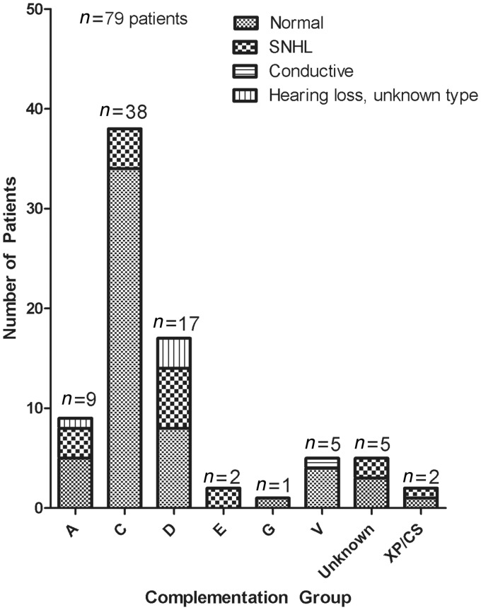
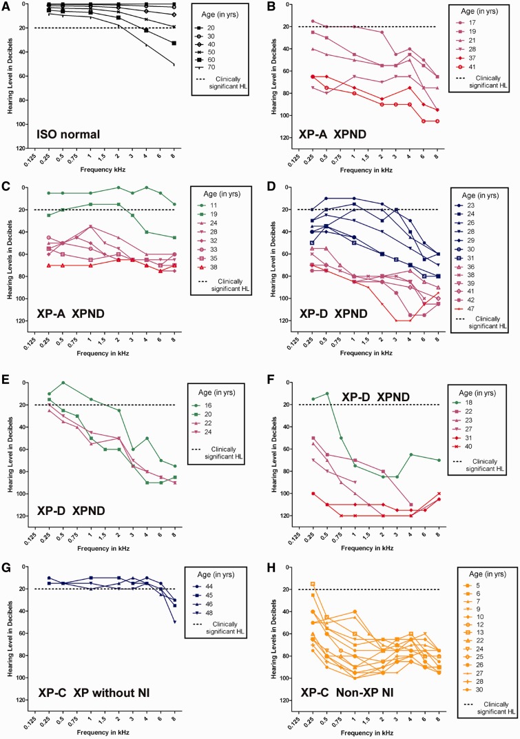
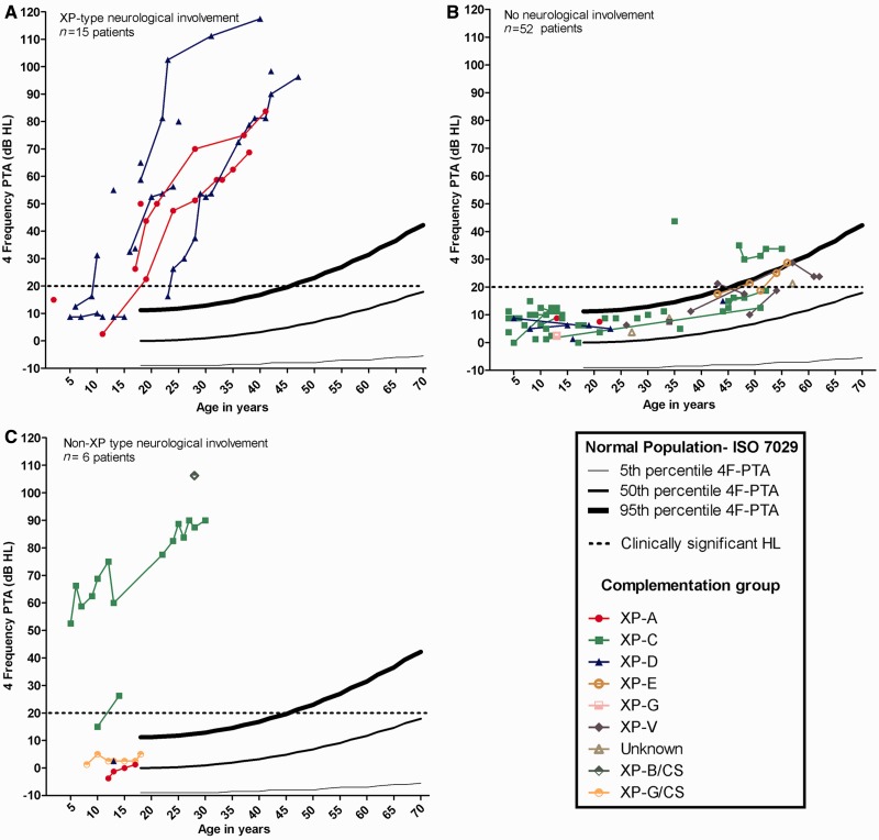
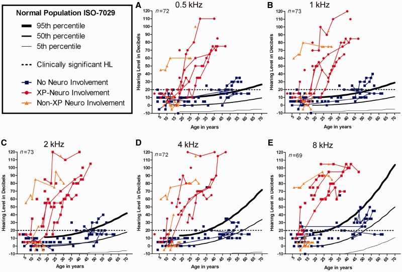
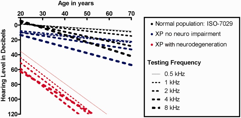
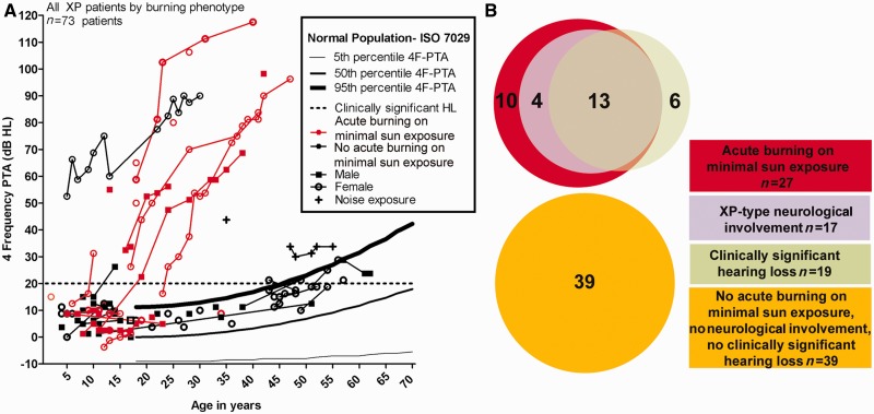
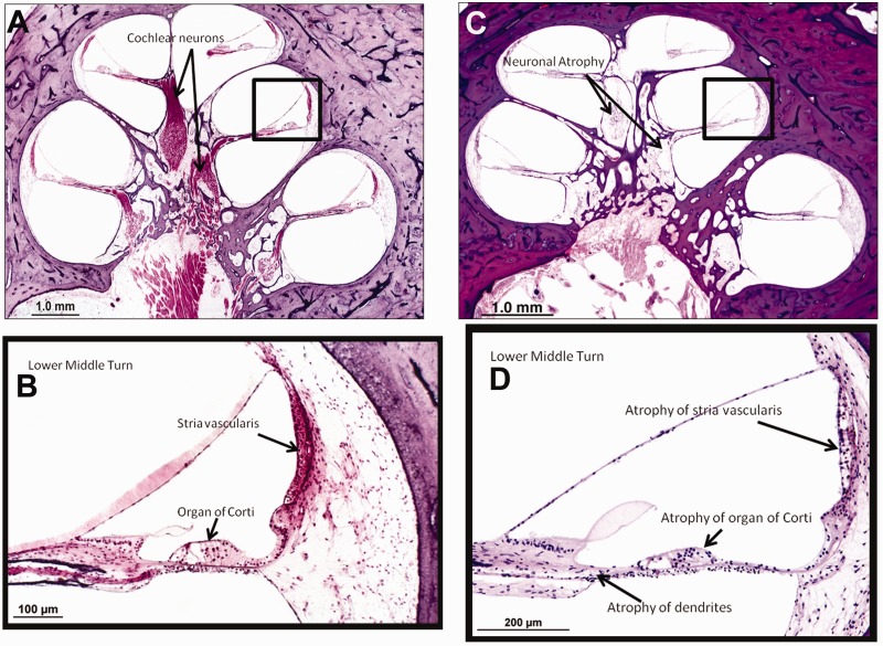
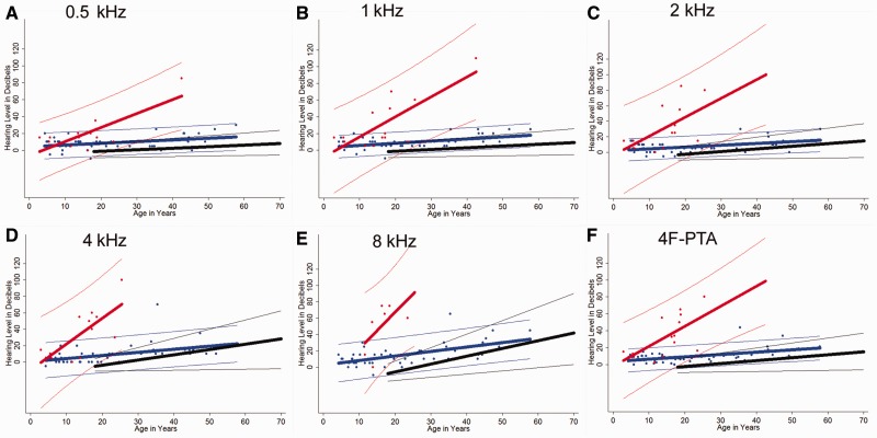
To assess the role of DNA repair in maintenance of hearing function and neurological integrity, we examined hearing status, neurological function, DNA repair complementation group and history of acute burning on minimal sun exposure in all patients with xeroderma pigmentosum, who had at least one complete audiogram, examined at the National Institutes of Health from 1971 to 2012. Seventy-nine patients, aged 1-61 years, were diagnosed with xeroderma pigmentosum (n = 77) or xeroderma pigmentosum/Cockayne syndrome (n = 2). A total of 178 audiograms were included. Clinically significant hearing loss (>20 dB) was present in 23 (29%) of 79 patients. Of the 17 patients with xeroderma pigmentosum-type neurological degeneration, 13 (76%) developed hearing loss, and all 17 were in complementation groups xeroderma pigmentosum type A or type D and reported acute burning on minimal sun exposure. Acute burning on minimal sun exposure without xeroderma pigmentosum-type neurological degeneration was present in 18% of the patients (10/55). Temporal bone histology in a patient with severe xeroderma pigmentosum-type neurological degeneration revealed marked atrophy of the cochlear sensory epithelium and neurons. The 19-year mean age of detection of clinically significant hearing loss in the patients with xeroderma pigmentosum with xeroderma pigmentosum-type neurological degeneration was 54 years younger than that predicted by international norms. The four frequency (0.5/1/2/4 kHz) pure-tone average correlated with degree of neurodegeneration (P < 0.001). In patients with xeroderma pigmentosum, aged 4-30 years, a four-frequency pure-tone average ≥10 dB hearing loss was associated with a 39-fold increased risk (P = 0.002) of having xeroderma pigmentosum-type neurological degeneration. Severity of hearing loss parallels neurological decline in patients with xeroderma pigmentosum-type neurological degeneration. Audiometric findings, complementation group, acute burning on minimal sun exposure and age were important predictors of xeroderma pigmentosum-type neurological degeneration. These results provide evidence that DNA repair is critical in maintaining neurological integrity of the auditory system.


Figures

Figure 1
Figure 1
Type of hearing loss by complementation group in patients with xeroderma pigmentosum seen at the National Institutes of Health from 1971 to 2012. Type of hearing loss is based on thresholds (dBHL) in the worse hearing ear at reference audiogram. XP/CS = xeroderma pigmentosum/Cockayne syndrome; SNHL = sensorineural hearing loss.
Figure 2
Figure 2
Longitudinal pure-tone air-conduction audiograms and degree of neurological involvement for individual patients with xeroderma pigmentosum. Individual patient thresholds for the worse hearing ear. Neurological status is colour coded: blue = no neurological involvement; green = mild mental retardation (IQ > 50), only hyporeflexia; purple = gait disturbance because of spasticity or ataxia, moderate mental retardation (IQ < 50); red = severe mental retardation, cannot walk, cannot speak and/or bed resting; orange = non-xeroderma pigmentosum-type neurological involvement [modified from Nishigori et al. (1994)]. (A) Age-based normative thresholds derived from combined male and female subject data from ISO 7029. (B) Patient XP12BE, XP-A with xeroderma pigmentosum-type neurological degeneration. (C) Patient XP19BE, XP-A with xeroderma pigmentosum-type neurological degeneration. (D) Patient XP33BE, XP-D with xeroderma pigmentosum-type neurological degeneration. (E) Patient XP29BE, XP-D with xeroderma pigmentosum-type neurological degeneration. (F) Patient XP18BE, XP-D with xeroderma pigmentosum-type neurological degeneration. (G) Patient XP-1BE, XP-C, xeroderma pigmentosum without neurological involvement (NI). (H) Patient XP21BE, XP-C with non-xeroderma pigmentosum type neurological involvement. HL = hearing loss; XP = xeroderma pigmentosum; XPND = xeroderma pigmentosum-type neurological degeneration.
Figure 3
Figure 3
Worse-hearing ear 4F-PTA plotted against the 95th, 50th and 5th percentiles obtained from age-matched normative data (ISO 7029) and the criterion for clinically normal hearing (20 dBHL). Data are plotted for all patients with xeroderma pigmentosum (XP) grouped by neurological status and colour-coded for xeroderma pigmentosum complementation group. (A) Patients with xeroderma pigmentosum-type neurological degeneration. (B) Patients with xeroderma pigmentosum without neurological involvement. The two XP-C patients above the 95th percentile range have documented noise exposure. (C) Patients with xeroderma pigmentosum with non-xeroderma pigmentosum-type neurological involvement. HL = hearing loss.
Figure 4
Figure 4
Worse-hearing ear pure-tone thresholds plotted against the percentiles obtained from age-matched normative data (ISO 7029) and the criterion for clinically normal hearing (20 dB hearing loss) for 0.5, 1, 2, 4 and 8 kHz. Neurological status, xeroderma pigmentosum-type neurological degeneration, patients with xeroderma pigmentosum (XP) without neurological involvement, and non-xeroderma pigmentosum type neurological involvement, is indicated by colour-coding. HL = hearing loss.
Figure 5
Figure 5
Linear regression analysis for age-based hearing decline at 0.5, 1, 2, 4 and 8 kHz. Regression is based on combined gender normative data (ISO 7029) and the worse hearing ear at the reference audiogram for patients with xeroderma pigmentosum-type neurological degeneration and patients with xeroderma pigmentosum without neurological involvement.
Figure 6
Figure 6
Acute burning on minimal sun exposure as an indicator of hearing loss (HL) and neurological involvement in patients with xeroderma pigmentosum. (A) Worse ear 4F-PTA in all patients with xeroderma pigmentosum grouped by acute burning on minimal sun exposure and plotted against the percentiles obtained from age-matched normative data (ISO 7029) and the criterion for clinically normal hearing (20 dB hearing loss). (B) Venn diagram showing acute burning on minimal sun exposure, hearing status and neurological involvement for patients with xeroderma pigmentosum with xeroderma pigmentosum-type neurological degeneration and no neurological involvement.
Figure 7
Figure 7
Cochlear histopathology in a patient with xeroderma pigmentosum-type neurodegeneration. (A) Cochlear histopathology of a normal-hearing individual at 63 years of age shown for comparison. (B) Higher power magnification of boxed area in (A). (C) Left ear cochlear pathology of Patient XP12BE (age 44 years) who developed xeroderma pigmentosum-type neurological degeneration. (D) Higher power magnification of boxed area in (C). Pathology shows diffuse atrophy of the organ of Corti, moderate to patchy atrophy of the stria vascularis and severe atrophy of cochlear neurons as compared with the control cochlea with normal hearing.
Figure 8
Figure 8
Regression curve analysis by Chow test. (AF) We compared combined gender ISO 7029 normative data (black), patients with xeroderma pigmentosum without neurological involvement (blue) and patients with xeroderma pigmentosum-type neurodegeneration (red) for 0.5, 1, 2, 4 and 8 kHz and the 4F-PTA. Regression curves for the patients with xeroderma pigmentosum are derived from the worse hearing ear at the earliest complete audiogram for each patient. The thicker line in the middle of each group of coloured data points represents a linear regression line corresponding to decibel level as predicted by age for that particular group. The thinner outer lines of each group mark the boundaries within which you would expect to find 95% of the sample’s values for a given age based on calculated prediction intervals. For the normal group, the recorded intervals represent those within which 95% of the population lies. P-values are <0.001 for all frequencies (including 4F-PTA) when comparing the combination of intercepts and slope for regression curves between xeroderma pigmentosum-type neurological degeneration, patients with xeroderma pigmentosum without neurological involvement and ISO normal populations, with the exception of ISO normal versus xeroderma pigmentosum no neurological involvement at 4 kHz (P = 0.006). All values are significant at Bonferroni-adjusted value of P < 0.002.

Similar articles

Cited by

References

    1. American National Standard Institute. New York: American National Standards Institute; 2003. Maximum permissible ambient noise levels for audiometric test rooms (Standard S3.1) (ANSI. S3.1-1999)
    1. American National Standards Institute. New York: American National Standards Institute; 2004. American national standard specification for audiometers (Standard S3.6) (ANSI. S3.6-1996)
    1. Anttinen A, Koulu L, Nikoskelainen E, Portin R, Kurki T, Erkinjuntti M, et al. Neurological symptoms and natural course of xeroderma pigmentosum. Brain. 2008;131:1979–89. - PubMed
    1. Bradford PT, Goldstein AM, Tamura D, Khan SG, Ueda T, Boyle J, et al. Cancer and neurologic degeneration in xeroderma pigmentosum: long term follow-up characterises the role of DNA repair. J Med Genet. 2011;48:168–76. - PMC - PubMed
    1. Brooks BP, Thompson AH, Clayton JA, Chan CC, Tamura D, Zein WM, et al. Ocular manifestations of trichothiodystrophy. Ophthalmology. 2011;118:2335–42. - PMC - PubMed

Publication types

MeSH terms