. 2013 Jan;136(Pt 1):223-44.
doi: 10.1093/brain/aws323.

New insights into genotype-phenotype correlations for the doublecortin-related lissencephaly spectrum

Collaborators, Affiliations

New insights into genotype-phenotype correlations for the doublecortin-related lissencephaly spectrum

Nadia Bahi-Buisson et al. Brain. 2013 Jan.

Abstract

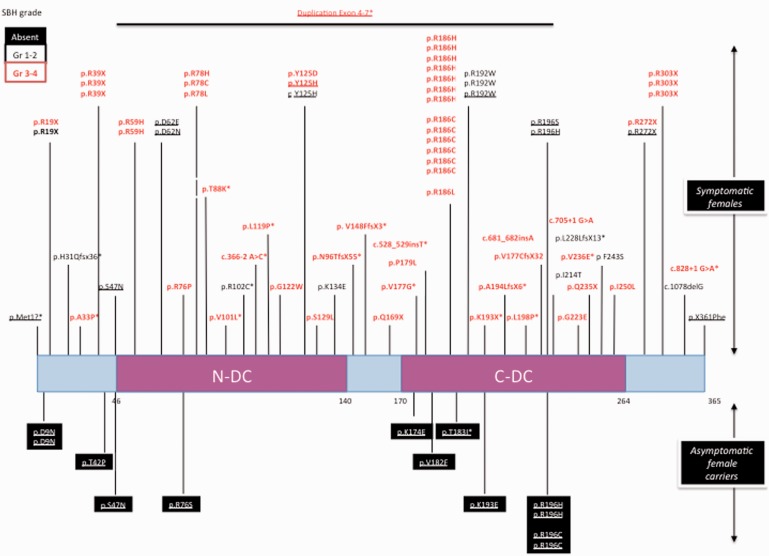
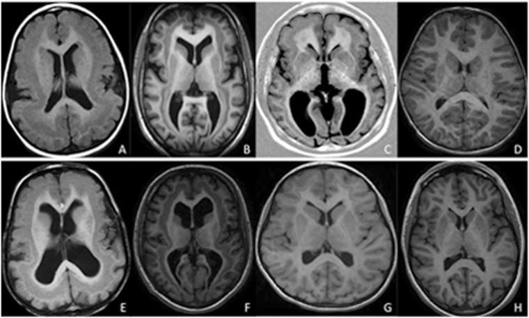
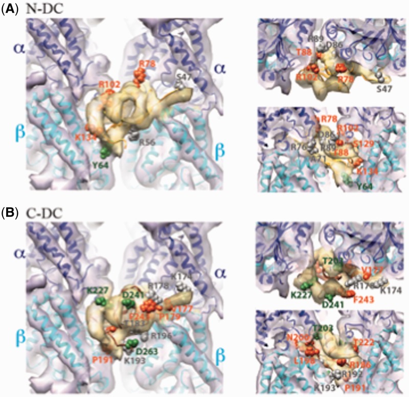
X-linked isolated lissencephaly sequence and subcortical band heterotopia are allelic human disorders associated with mutations of doublecortin (DCX), giving both familial and sporadic forms. DCX encodes a microtubule-associated protein involved in neuronal migration during brain development. Structural data show that mutations can fall either in surface residues, likely to impair partner interactions, or in buried residues, likely to impair protein stability. Despite the progress in understanding the molecular basis of these disorders, the prognosis value of the location and impact of individual DCX mutations has largely remained unclear. To clarify this point, we investigated a cohort of 180 patients who were referred with the agyria-pachygyria subcortical band heterotopia spectrum. DCX mutations were identified in 136 individuals. Analysis of the parents' DNA revealed the de novo occurrence of DCX mutations in 76 cases [62 of 70 females screened (88.5%) and 14 of 60 males screened (23%)], whereas in the remaining cases, mutations were inherited from asymptomatic (n = 14) or symptomatic mothers (n = 11). This represents 100% of families screened. Female patients with DCX mutation demonstrated three degrees of clinical-radiological severity: a severe form with a thick band (n = 54), a milder form (n = 24) with either an anterior thin or an intermediate thickness band and asymptomatic carrier females (n = 14) with normal magnetic resonance imaging results. A higher proportion of nonsense and frameshift mutations were identified in patients with de novo mutations. An analysis of predicted effects of missense mutations showed that those destabilizing the structure of the protein were often associated with more severe phenotypes. We identified several severe- and mild-effect mutations affecting surface residues and observed that the substituted amino acid is also critical in determining severity. Recurrent mutations representing 34.5% of all DCX mutations often lead to similar phenotypes, for example, either severe in sporadic subcortical band heterotopia owing to Arg186 mutations or milder in familial cases owing to Arg196 mutations. Taken as a whole, these observations demonstrate that DCX-related disorders are clinically heterogeneous, with severe sporadic and milder familial subcortical band heterotopia, each associated with specific DCX mutations. There is a clear influence of the individual mutated residue and the substituted amino acid in determining phenotype severity.


Figures

Figure 1
Figure 1
Schematic representation of the DCX protein and summary of the mutations identified in asymptomatic and symptomatic females with SBH. Each DCX nucleotide mutation is numbered with reference to the ATG. The predicted DCX protein alteration is numbered with reference to the amino acid (aa) residue number. Amino acid substitution mutations are referenced by the wild-type amino acid and position followed by the mutant amino acid. For example, D9N indicates that the wild-type amino acid D at position 9 is mutated to an N. One or two base-pair deletions or insertions result in a translational reading frameshift followed by a protein termination codon. Mutations are indicated on the protein sequence of DCX that comprises two evolutionary conserved domains clustered in two repeats, namely, N-DC aa 46–139 and C-DC aa 173–263 depicted as pink boxes, and the N-terminal, the interdomain and the C-terminal linker are shown as light blue boxes. The mutations found in symptomatic females are indicated above the gene, and the deleted portions of the gene as thick lines and mutations found in asymptomatic females are shown in black boxes below the gene schema. An asterisk denotes the mutations described in this article. Severe forms with SBH grade 3–4 are denoted in red, and milder forms with SBH grade 1–2 are denoted in black. Inherited mutations are underlined.
Figure 2
Figure 2
Schematic representation of the DCX protein and summary of the mutations identified in males with lissencephaly. Each DCX nucleotide mutation is numbered with reference to the ATG. The predicted DCX protein alteration is numbered with reference to the amino acid (aa) residue number. Mutations are indicated on the protein sequence of DCX that comprises two evolutionary conserved domains clustered in two repeats, namely, N-DC aa 46–139 and C-DC aa 173–263 depicted as pink boxes, and the N-terminal, the interdomain and the C-terminal linker are shown as light blue boxes. The inherited mutations are indicated above the gene, the families with two brothers affected are figures in black boxes, and de novo mutations found are shown below the gene scheme and the deleted portions of the gene as thick lines. An asterisk denotes the mutations described in this article. Severe forms with LIS grade 1–2 are denoted in red, intermediate forms with LIS grade 3–4 are denoted in black, and in light grey boxes are the milder forms with LIS grade 5–6. Inherited mutations are underlined.
Figure 3
Figure 3
Variable extent and thickness of band in cases with sporadic SBH. Representative MRI scans in patients with either a thick continuous band around the entire brain (grade 3–4) (A, B, E and F) or a thinner band only present in the frontal lobe (G) or restricted to the frontal and temporal lobe with intermediate thickness (grade 2) (C). Thick and continuous band around the entire brain in two patients aged 2 years and 11 months (A) and 3 years and 6 months (E), respectively. The bands appear to fuse with the outer cortex in the frontal regions. Intermediate diffuse SBH in two patients aged 15 (B) and 16 years (F). Thin band only present in the frontal lobe (G) or restricted to the frontal and the temporal lobe (C) in two patients aged 24 years and 11 years. D and H are from control patients aged 3 and 15 years, respectively.
Figure 4
Figure 4
Representative axial MRI in young children aged 7 months (A and E), 14 months (B and F) and 17 months (C and G). T1-weighted images show poor differentiation of the cortex and underlying white matter, with an aspect reminiscent of diffuse pachygyria (in younger child, A), or frontoparietal pachygyria combined with band heterotopia in posterior regions (in patients >1 year of age, B and C). At the same level, on T2-weighted images, the band is visible (E, F and G). Control MRI: T1-weighted image (D) and T2-weighted image (H) in a normal 15-month-old female.
Figure 5
Figure 5
Representative T1-weighted (A, B and C) and T2-weighted (E, F and G) axial section of MRI in three males with DCX mutations representing the most prominent LIS grade in this study. Anterior pachygyria (LIS grade 4) in a 2-year-old male (familial case) (A and E). SBH with anterior pachygyria in a 5-year-old male (familial case) (B and F). Severe lissencephaly (LIS grade 2) more severe anteriorly in a sporadic male aged 1 year 3 months (C and G). Control MRI: T1-weighted image (D) and T2-weighted image (H) in normal 18-month-old boy.
Figure 6
Figure 6
Localization of surface residues mutated in DCX in SBH and their relationship with the microtubule interface. (A) Structure of the DCX–microtubule interface [cryo-electron microscopy reconstruction displayed as a transparent surface, tubulin in purple, DCX in yellow; EMDB ID 1788 (Kim et al., 2003; Fourniol et al., 2010)] docked with the pseudo-atomic structure of the N-DC–microtubule interface [ribbons, α-tubulin in blue, β-tubulin in cyan, N-DC in orange; PDB ID 2XRP (Kim et al., 2003; Fourniol et al., 2010)]. Left: front view; top right: view from the microtubule plus end; bottom right: view from the centre of the microtubule outwards. N-DC surface residues subject to missense mutations are displayed as spheres, coloured in green for cases with absence of cortex malformations (R102, K134), grey for mild/moderate phenotypes (S47), and orange for severe cases (R59, Y64, R76, R78, S129). Note that when a mutation resulted in more or less severe SBH in different patients, the most severe phenotype was considered in this figure. (B) Same as in A but docked with a homology model of C-DC (ribbons, brown). Green spheres: surface residue, the mutation of which resulted in an absence of phenotype (K193; however, X-inactivation status for the individual with this mutation is not available, and this residue results in a severe phenotype in a male with lissencephaly); grey spheres: surface residues the mutation of which caused milder SBH (K174, T183, R196, T203, K227, F243 and D263); orange: severe SBH cases (V177, P179, R186, R192, L198 and D241). Of note, milder-effect mutations apparently appear more frequently in C-DC than in N-DC.

Similar articles

Cited by

References

    1. Abdollahi MR, Morrison E, Sirey T, Molnar Z, Hayward BE, Carr IM, et al. Mutation of the variant alpha-tubulin TUBA8 results in polymicrogyria with optic nerve hypoplasia. Am J Hum Genet. 2009;85:737–44. - PMC - PubMed
    1. Ackman JB, Aniksztejn L, Crepel V, Becq H, Pellegrino C, Cardoso C, et al. Abnormal network activity in a targeted genetic model of human double cortex. J Neurosci. 2009;29:313–27. - PMC - PubMed
    1. Aigner L, Fluegel D, Dietrich J, Ploetz S, Winkler J. Isolated lissencephaly sequence and double-cortex syndrome in a German family with a novel doublecortin mutation. Neuropediatrics. 2000;31:195–8. - PubMed
    1. Aigner L, Uyanik G, Couillard-Despres S, Ploetz S, Wolff G, Morris-Rosendahl D, et al. Somatic mosaicism and variable penetrance in doublecortin-associated migration disorders. Neurology. 2003;60:329–32. - PubMed
    1. Barkovich AJ, Guerrini R, Battaglia G, Kalifa G, N'Guyen T, Parmeggiani A, et al. Band heterotopia: correlation of outcome with magnetic resonance imaging parameters. Ann Neurol. 1994;36:609–17. - PubMed

Publication types

MeSH terms