

Rod photoreceptors protect from cone degeneration-induced retinal remodeling and restore visual responses in zebrafish
- PMID: 23365220
- PMCID: PMC3711385
- DOI: 10.1523/JNEUROSCI.2910-12.2013
Rod photoreceptors protect from cone degeneration-induced retinal remodeling and restore visual responses in zebrafish
Abstract
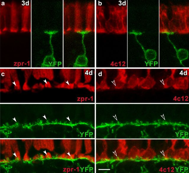
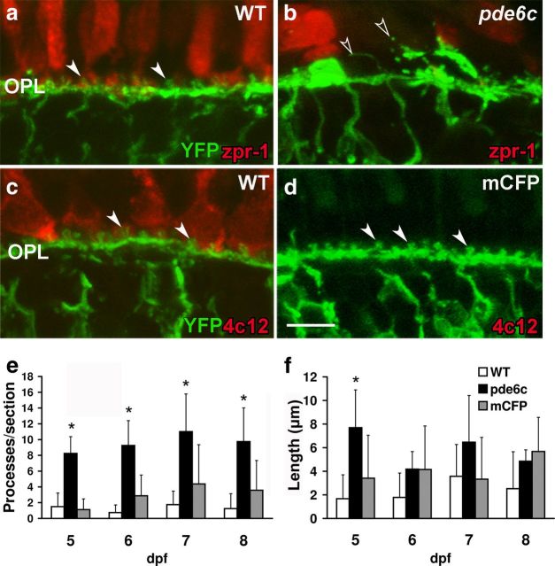
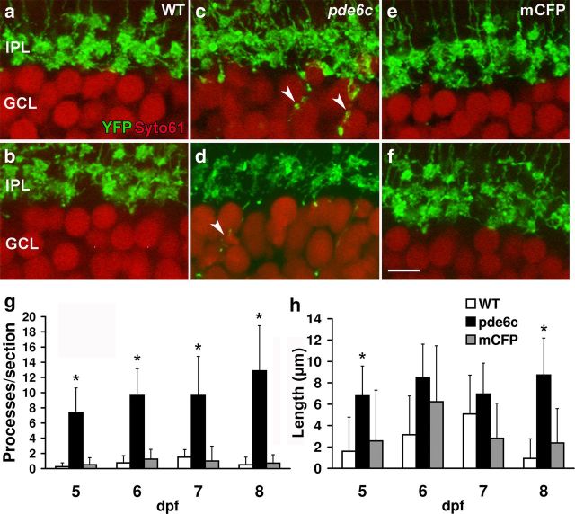
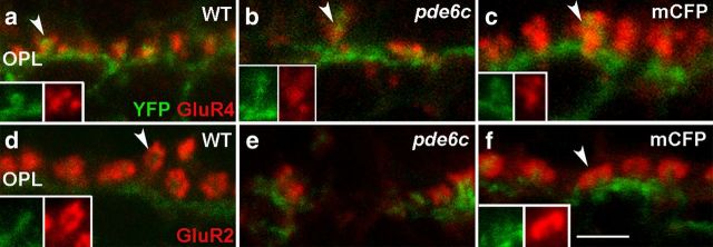
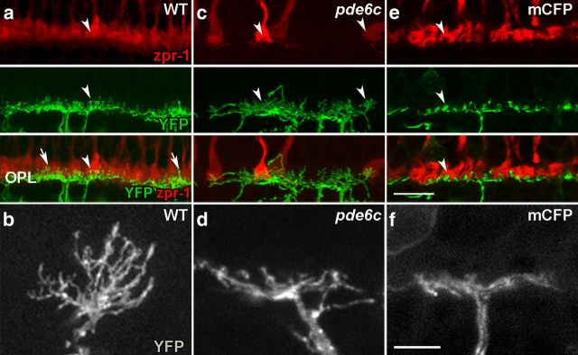
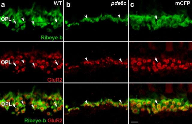
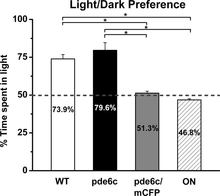
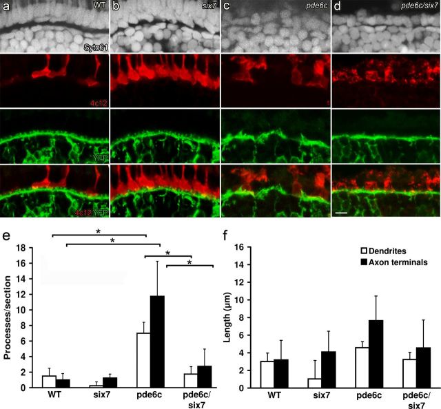
Humans are largely dependent upon cone-mediated vision. However, death or dysfunction of rods, the predominant photoreceptor subtype, results in secondary loss of cones, remodeling of retinal circuitry, and blindness. The changes in circuitry may contribute to the vision deficit and undermine attempts at restoring sight. We exploit zebrafish larvae as a genetic model to specifically characterize changes associated with photoreceptor degenerations in a cone-dominated retina. Photoreceptors form synapses with two types of second-order neurons, bipolar cells, and horizontal cells. Using cell-specific reporter gene expression and immunolabeling for postsynaptic glutamate receptors, significant remodeling is observed following cone degeneration in the pde6c(w59) larval retina but not rod degeneration in the Xops:mCFP(q13) line. In adults, rods and cones are present in approximately equal numbers, and in pde6c(w59) mutants glutamate receptor expression and synaptic structures in the outer plexiform layer are preserved, and visual responses are gained in these once blind fish. We propose that the abundance of rods in the adult protects the retina from cone degeneration-induced remodeling. We test this hypothesis by genetically manipulating the number of rods in larvae. We show that an increased number and uniform distribution of rods in lor/tbx2b(p25bbtl) or six7 morpholino-injected larvae protect from pde6c(w59)-induced secondary changes. The observations that remodeling is a common consequence of photoreceptor death across species, and that in zebrafish a small number of surviving photoreceptors afford protection from degeneration-induced changes, provides a model for systematic analysis of factors that slow or even prevent the secondary deteriorations associated with neural degenerative disease.
Figures
Similar articles
-
Cone survival despite rod degeneration in XOPS-mCFP transgenic zebrafish.Invest Ophthalmol Vis Sci. 2005 Dec;46(12):4762-71. doi: 10.1167/iovs.05-0797. Invest Ophthalmol Vis Sci. 2005. PMID: 16303977 Free PMC article.
-
Ablation of retinal horizontal cells from adult mice leads to rod degeneration and remodeling in the outer retina.J Neurosci. 2012 Aug 1;32(31):10713-24. doi: 10.1523/JNEUROSCI.0442-12.2012. J Neurosci. 2012. PMID: 22855819 Free PMC article.
-
Structural and functional remodeling in the retina of a mouse with a photoreceptor synaptopathy: plasticity in the rod and degeneration in the cone system.Eur J Neurosci. 2007 Nov;26(9):2506-15. doi: 10.1111/j.1460-9568.2007.05886.x. Epub 2007 Oct 23. Eur J Neurosci. 2007. PMID: 17970721
-
Neural remodeling in retinal degeneration.Prog Retin Eye Res. 2003 Sep;22(5):607-55. doi: 10.1016/s1350-9462(03)00039-9. Prog Retin Eye Res. 2003. PMID: 12892644 Review.
-
Therapeutic strategy for handling inherited retinal degenerations in a gene-independent manner using rod-derived cone viability factors.C R Biol. 2014 Mar;337(3):207-13. doi: 10.1016/j.crvi.2013.12.002. Epub 2014 Feb 20. C R Biol. 2014. PMID: 24702847 Review.
Cited by
-
Targeted disruption of the endogenous zebrafish rhodopsin locus as models of rapid rod photoreceptor degeneration.Mol Vis. 2018 Aug 27;24:587-602. eCollection 2018. Mol Vis. 2018. PMID: 30210230 Free PMC article.
-
Defects in the retina of Niemann-pick type C 1 mutant mice.BMC Neurosci. 2014 Nov 29;15:126. doi: 10.1186/s12868-014-0126-2. BMC Neurosci. 2014. PMID: 25472750 Free PMC article.
-
A Comparative Transcriptomic Analysis of Development in Two Astyanax Cavefish Populations.J Exp Zool B Mol Dev Evol. 2017 Sep;328(6):515-532. doi: 10.1002/jez.b.22749. Epub 2017 Jun 14. J Exp Zool B Mol Dev Evol. 2017. PMID: 28612405 Free PMC article.
-
Genetic Dissection of Dual Roles for the Transcription Factor six7 in Photoreceptor Development and Patterning in Zebrafish.PLoS Genet. 2016 Apr 8;12(4):e1005968. doi: 10.1371/journal.pgen.1005968. eCollection 2016 Apr. PLoS Genet. 2016. PMID: 27058886 Free PMC article.
-
Mutation of wrb, a Component of the Guided Entry of Tail-Anchored Protein Pathway, Disrupts Photoreceptor Synapse Structure and Function.Invest Ophthalmol Vis Sci. 2016 Jun 1;57(7):2942-54. doi: 10.1167/iovs.15-18996. Invest Ophthalmol Vis Sci. 2016. PMID: 27273592 Free PMC article.
References
-
- Ball SL, Pardue MT, McCall MA, Gregg RG, Peachey NS. Immunohistochemical analysis of the outer plexiform layer in the nob mouse shows no abnormalities. Vis Neurosci. 2003;20:267–272. - PubMed
-
- Beck JC, Gilland E, Tank DW, Baker R. Quantifying the ontogeny of optokinetic and vestibuloocular behaviors in zebrafish, medaka, and goldfish. J Neurophysiol. 2004;92:3546–3561. - PubMed
Publication types
MeSH terms
Substances
Grants and funding
LinkOut - more resources
Full Text Sources
Other Literature Sources
Molecular Biology Databases