. 2013 Jan 30;33(5):1991-2008.
doi: 10.1523/JNEUROSCI.0541-12.2013.

Where one hand meets the other: limb-specific and action-dependent movement plans decoded from preparatory signals in single human frontoparietal brain areas

Affiliations

Where one hand meets the other: limb-specific and action-dependent movement plans decoded from preparatory signals in single human frontoparietal brain areas

Jason P Gallivan et al. J Neurosci. .

Abstract

Planning object-directed hand actions requires successful integration of the movement goal with the acting limb. Exactly where and how this sensorimotor integration occurs in the brain has been studied extensively with neurophysiological recordings in nonhuman primates, yet to date, because of limitations of non-invasive methodologies, the ability to examine the same types of planning-related signals in humans has been challenging. Here we show, using a multivoxel pattern analysis of functional MRI (fMRI) data, that the preparatory activity patterns in several frontoparietal brain regions can be used to predict both the limb used and hand action performed in an upcoming movement. Participants performed an event-related delayed movement task whereby they planned and executed grasp or reach actions with either their left or right hand toward a single target object. We found that, although the majority of frontoparietal areas represented hand actions (grasping vs reaching) for the contralateral limb, several areas additionally coded hand actions for the ipsilateral limb. Notable among these were subregions within the posterior parietal cortex (PPC), dorsal premotor cortex (PMd), ventral premotor cortex, dorsolateral prefrontal cortex, presupplementary motor area, and motor cortex, a region more traditionally implicated in contralateral movement generation. Additional analyses suggest that hand actions are represented independently of the intended limb in PPC and PMd. In addition to providing a unique mapping of limb-specific and action-dependent intention-related signals across the human cortical motor system, these findings uncover a much stronger representation of the ipsilateral limb than expected from previous fMRI findings.


Figures

Figure 1.
Figure 1.
Experimental methods. A, Subject setup from side view (note that the bore cameras are not shown). B, Experimental apparatus and target object shown from the subject's point of view (POV). The target object (centrally located) never changed position from trial to trial. Green star with dark shadow represents the fixation LED and its location in depth. The left and right hands are positioned at their respective starting positions. C, Executed hand movements. D, Timing of one event-related trial. Trials began with the 3D target being illuminated while the subject maintained fixation (preview phase; 6 s). Participants were then instructed via headphones to perform one of four movements: grasp with the left hand (“grasp left”), grasp with the right hand (“grasp right”), reach with the left hand (“reach left”), or reach with the right hand (“reach right”). This cue initiated the plan phase portion of the trial. After a fixed delay interval (12 s), participants were then cued (“Beep”) to perform the instructed hand movement (initiating the execute phase portion of the trial). Two seconds after the go cue, vision of the workspace was extinguished, cuing participants to return their hand to its starting position and then wait for the following trial to begin (14 s, ITI). E, Averaged neural activity from the left SMA (L-SMA) over the length of a single trial. Events in E are time-locked to correspond to events in D. MVPA was performed on single trials based on the windowed average of the percentage signal change response corresponding to the three different time epochs denoted by each of the gray shaded bars (each corresponding to activity elicited from the three distinct trial phases: preview, plan, and execute). To examine what types of upcoming movements could be predicted, decoding brain activity from the premovement time points (bordered in blue) was of critical interest.
Figure 2.
Figure 2.
Decoding of limb-specific and action-dependent intention-related signals across frontoparietal cortex. Cortical areas that exhibited larger responses during movement generation than the preceding visual phase [execute > preview] are shown in orange/yellow activation (see statistical thresholds, at bottom). Results calculated across all participants (RFX GLM) are displayed on one representative subject's inflated hemispheres. The general locations of the selected ROIs are outlined in circles (actual ROIs were anatomically defined separately in each subject). The outline of each ROI is color coded according to the general pattern of pairwise discriminations made during movement planning; see color legend at top for classification profiles (for reference, see Figs. 3–8). Colors pertain to significant decoding accuracies for plan phase preparatory activity with respect to 50% chance classification (with respect to the black asterisks in Figs. 3–8). Sulcal landmarks are denoted by white lines (stylized according to the corresponding legend shown at bottom). LH, Left hemisphere; RH, right hemisphere.
Figure 3.
Figure 3.
Movement plan decoding in PPC. Each individual ROI is associated with three plots of data. Top, Percentage signal change time course activity. The activity in each plot is averaged across all voxels within each ROI and across participants. Vertical lines correspond to the onset of the preview, plan, and execute phases of each trial (from left to right). Shaded gray bars indicate the 2-volume (4 s) windows that were averaged and extracted for MVPA. Time corresponds to seconds. Bottom left, Corresponding decoding accuracies are shown for each time phase (preview, plan, and execute). Classifier training and testing was done using a single trial N − 1 cross-validation procedure. Note that accurate classification is primarily attributable to the spatial activity patterns of different planned movement types and not to differences in the overall signal amplitude responses within each ROI (i.e., time courses are highly overlapping during the plan phase). Bottom right, Cross-decoding accuracies are shown for each time phase (preview, plan, and execute). Limb-specific, action-independent accuracies were computed from training classifiers on GraspL versus GraspR trials and testing on ReachL versus ReachR trials and then averaging the resulting accuracies with those obtained from the opposite train-and-test ordering, within each subject. Action-specific, limb-independent accuracies were computed from training classifiers on GraspL versus ReachL trials and testing on GraspR versus ReachR trials (again, averaging these resulting accuracies with those obtained from the opposite train-and-test ordering, within each subject). Error bars represent SEM across participants. Solid black lines are chance accuracy level (50%). Black asterisks assess statistical significance with two-tailed t tests across participants with respect to 50%. Red asterisks assess statistical significance based on an FDR correction of q ≤ 0.05. For further details, see Materials and Methods.
Figure 4.
Figure 4.
Movement plan decoding in anterior parietal cortex. Percentage signal change time courses and decoding accuracies are plotted and computed the same as in Figure 3.
Figure 5.
Figure 5.
Movement plan decoding in motor and SS cortices. Percentage signal change time courses and decoding accuracies are plotted and computed the same as in Figure 3. Note the significant decoding in motor cortex during the plan phase despite near baseline-level activity in the percentage signal change responses. Also note that significant decoding in SS cortex only arises after movement onset (i.e., execute phase), once the mechanoreceptors of the hand have been stimulated by object contact.
Figure 6.
Figure 6.
Movement plan decoding in premotor cortex. Percentage signal change time courses and decoding accuracies are plotted and computed the same as in Figure 3.
Figure 7.
Figure 7.
Movement plan decoding in the SMAs. Percentage signal change time courses and decoding accuracies are plotted and computed the same as in Figure 3.
Figure 8.
Figure 8.
Movement plan decoding in prefrontal cortex. Percentage signal change time courses and decoding accuracies are plotted and computed the same as in Figure 3.
Figure 9.
Figure 9.
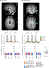
Decoding in the non-brain control ROIs. Non-brain control ROIs defined in each subject (denoted in cyan; example subject shown). Percentage signal change time courses and decoding accuracies are plotted and computed the same as in Figure 3. Note that no significant differences were found with t tests across participants with respect to 50% chance. A, Anterior; P, posterior.

References

    1. Aizawa H, Mushiake H, Inase M, Tanji J. An output zone of the monkey primary motor cortex specialized for bilateral hand movement. Exp Brain Res. 1990;82:219–221. - PubMed
    1. Andersen RA, Buneo CA. Intentional maps in posterior parietal cortex. Annu Rev Neurosci. 2002;25:189–220. - PubMed
    1. Andersen RA, Cui H. Intention, action planning, and decision making in parietal-frontal circuits. Neuron. 2009;63:568–583. - PubMed
    1. Barash S, Bracewell RM, Fogassi L, Gnadt JW, Andersen RA. Saccade-related activity in the lateral intraparietal area. I. Temporal properties; comparison with area 7a. J Neurophysiol. 1991;66:1095–1108. - PubMed
    1. Basso MA, Wurtz RH. Modulation of neuronal activity by target uncertainty. Nature. 1997;389:66–69. - PubMed

Publication types

LinkOut - more resources