. 2013 Apr;87(7):4060-70.
doi: 10.1128/JVI.02020-12. Epub 2013 Jan 30.

Epstein-Barr virus deubiquitinase downregulates TRAF6-mediated NF-κB signaling during productive replication

Affiliations

Epstein-Barr virus deubiquitinase downregulates TRAF6-mediated NF-κB signaling during productive replication

Shinichi Saito et al. J Virol. 2013 Apr.

Abstract

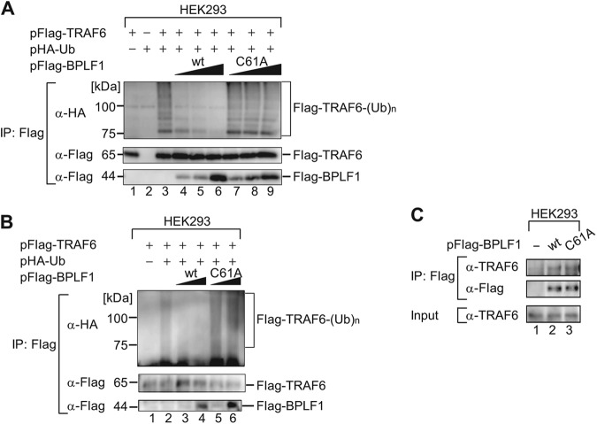
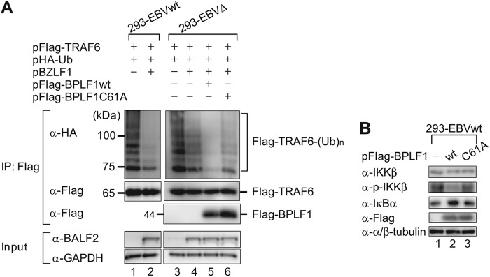
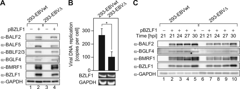
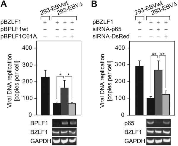
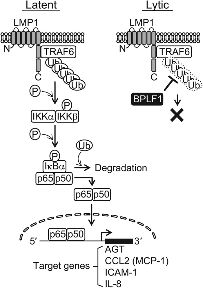
Epstein-Barr virus (EBV), a human oncogenic herpesvirus that establishes a lifelong latent infection in the host, occasionally enters lytic infection to produce progeny viruses. The EBV oncogene latent membrane protein 1 (LMP1), which is expressed in both latent and lytic infection, constitutively activates the canonical NF-κB (p65) pathway. Such LMP1-mediated NF-κB activation is necessary for proliferation of latently infected cells and inhibition of viral lytic cycle progression. Actually, canonical NF-κB target gene expression was suppressed upon the onset of lytic infection. TRAF6, which is activated by conjugation of polyubiquitin chains, associates with LMP1 to mediate NF-κB signal transduction. We have found that EBV-encoded BPLF1 interacts with and deubiquitinates TRAF6 to inhibit NF-κB signaling during lytic infection. HEK293 cells with BPLF1-deficient recombinant EBV exhibited poor viral DNA replication compared with the wild type. Furthermore, exogenous expression of BPLF1 or p65 knockdown in cells restored DNA replication of BPLF1-deficient viruses, indicating that EBV BPLF1 deubiquitinates TRAF6 to inhibit NF-κB signal transduction, leading to promotion of viral lytic DNA replication.


Figures

Fig 1
Fig 1
Ectopic expression of BPLF1 decreases NF-κB-dependent promoter activity in cells latently infected with EBV. Latently infected B95-8, AGS-EBV, and 293-EBVwt cells were transfected with the NF-κB-Fluc reporter plasmid (0.2 μg/well), along with the pCMV-Rluc plasmid (0.02 μg/well) and either pBPLF1wt or pBPLF1C61A (0.1 μg/well), in 24-well plates. Luciferase assays were performed at 24 hpt. Firefly luciferase activity was normalized to Renilla reniformis luciferase, and the value obtained by transfecting an empty-vector control was set to 100%. Data are shown as means ± SD of the results of 3 biological replicates. **, P < 0.001; *, P < 0.005. Sample lysates were subsequently subjected to immunoblotting with the specific antibodies indicated, and representative results are presented below the graph. In addition, sample lysates of cells transfected with BZLF1 were also included as controls for lytic replication.
Fig 2
Fig 2
Recombinant EBV-BAC genome structures. (A) Schematic arrangement of recombination of the EBV genome using the neomycin resistance and streptomycin sensitivity genes. The region between nucleotides 1 and 975 of the BPLF1 ORF was replaced with tandemly arranged neomycin resistance and streptomycin sensitivity (NeoSt+) genes to make dBPLF1/NeoSt. (B and C) Electrophoresis of wild-type and recombinant EBV-BAC DNAs. EBV-BAC DNAs were digested with BamHI (B) or EcoRI (C) and separated in a 0.8% agarose gel. The sizes of BamHI-P fragment and a corresponding EcoRI fragment of the EBV-BAC DNAs (open arrows) were shifted by integration of the marker cassettes (closed arrows). Sizes (kbp) for molecular mass markers are indicated at the left side of the panels. (D) PCR analysis of the wild-type and the recombinant BAC DNAs with BPLF1 ORF-specific primers. The PCR product was detected by 1.5% agarose gel electrophoresis. (E) RT-PCR analysis of BPLF1 expressed in pBZLF1-transfected 293-EBVwt and 293-EBVΔ cells. Total RNAs were extracted at 48 hpi, and cDNAs were synthesized as described in Materials and Methods. PCR was performed on cDNA templates with specific primers. BZLF1 was used as an induction marker and GAPDH as an internal control. (F) Total DNAs prepared from 293-EBVwt and 293-EBVΔ cells were applied to qrt-PCR using BALF2-specific primers to quantify intracellular EBV-BAC DNA copies. The values were normalized to that of Namalwa cells, which maintain 2 EBV genomes per cell. (G) Western blotting using anti-LMP1 antibody was performed using whole-cell lysate prepared from 293-EBVwt and 293-EBVΔ cells to confirm that comparable amounts of the latent protein are expressed in both cells. (H) 293-EBVwt cells transfected with 1 μg of pBZLF1 were cultured for indicated periods, and expression levels of BPLF1 mRNA were measured by RT-PCR. Three biological replicates were carried out for the time-course analysis. Data from one representative experiment are shown.
Fig 3
Fig 3
DUB activity is essential for BPLF1 to block activation of the NF-κB pathway during EBV lytic replication. Wild-type and recombinant BPLF1 expression vectors (0.1 μg each) were cotransfected into 293-EBVwt and 293-EBVΔ cells along with the pBZLF1 plasmid (1 μg), the pNF-κB-Fluc reporter plasmid (0.2 μg), and pCMV-Rluc (0.02 μg) using an MP-100 electroporator. An empty vector (pcDNA3) was used as a control. Cell extracts were collected at 24 hpt and analyzed for firefly and Renilla luciferase expression. Firefly luciferase activity was normalized to the Renilla reniformis luciferase, and the values obtained by transfecting the empty-plasmid control into 293-EBVwt or 293-EBVΔ were set to 100%. Sample lysates were subsequently subjected to immunoblotting with specific antibodies, and a representative result is presented below the graph. BZLF1 (immediate-early) and BALF2 (early) were used as induction markers. Data are shown as means ± SD of the results of 5 biological replicates. ***, P < 0.001; **, P < 0.005; *, P < 0.01.
Fig 4
Fig 4
BPLF1 suppresses expression of NF-κB-regulated genes during the EBV lytic life cycle. (A) 293-EBVwt and 293-EBVΔ cells were transfected with control or BZLF1 expression plasmids. At 24 hpi, cells were subjected to qrt-PCR to measure the mRNA levels of NF-κB-dependent genes. Values were normalized to GAPDH mRNA, and the value obtained by transfecting an empty-plasmid control into 293-EBVwt or 293-EBVΔ was set to 1. Data are shown as means ± SD of the results of 3 biological replicates. ***, P < 0.001; **, P < 0.005; *, P < 0.05. (B) RT-PCR was carried out in order to detect BPLF1 mRNA in the same samples described for panel A, followed by an agarose electrophoresis.
Fig 5
Fig 5
BPLF1 interacts with and inhibits ubiquitination of TRAF6. (A) HEK293 cells cultured in 6-well plates were cotransfected with hemagglutinin (HA)-tagged Ub (2 μg/well) and TRAF6 (3 μg/well) expression plasmids and increasing quantities (0.1, 0.2, or 0.5 μg/well) of the designated BPLF1 expression plasmid. Cell lysates were prepared at 24 hpi and immunoprecipitated (IP) with anti-Flag antibodies, and ubiquitin conjugation of the TRAF6 protein was verified by immunoblotting with anti-HA antibodies. Production of exogenously expressed tagged proteins was verified with the indicated antibodies. The experiment shown is a representative of three independent experiments. (B) The conditions were basically the same as described for panel A except that cells were lysed with the denaturing lysis buffer containing 2% SDS followed by a 10-min incubation at 95°C. The amount of transfected BPLF1 expression plasmid was 0.1 or 0.5 μg/well. The experiment shown is a representative of three independent experiments. (C) HEK293 cells cultured in 6-well plates were transfected with an empty plasmid or designated BPLF1 (0.5 μg/well) expression plasmids. Cell lysates were prepared at 24 hpi and immunoprecipitated with anti-Flag antibodies, followed by immunoblot analysis with anti-TRAF6 antibodies. Production of exogenously expressed BPLF1 proteins was verified with anti-Flag antibody. The experiment shown is a representative of two independent experiments.
Fig 6
Fig 6
Endogenous BPLF1 deubiquitinates TRAF6. (A) 293-EBVwt and 293-EBVΔ cells cultured in 6-well plates were cotransfected with HA-tagged Ub (2 μg/well), TRAF6 (3 μg/well), and BZLF1 (1 μg/well) expression plasmids. Four hours after the initial transfection, the cells were further transfected with either wild-type or enzyme-dead BPLF1 expression plasmids (0.5 μg/well). Cell lysates were prepared at 24 h after initial transfection, and immunoprecipitation experiments were performed in the same fashion as described for Fig. 5A. The experiment shown is a representative of three independent experiments. (B) 293-EBVwt cells were transfected with empty plasmid or designated BPLF1 (0.5 μg/well) expression plasmid. Cell lysates were prepared at 24 hpi, and immunoblot analysis was performed using indicated antibodies. The experiment shown is a representative of two independent experiments.
Fig 7
Fig 7
BPLF1 promotes EBV genome replication. (A) 293-EBVwt and 293-EBVΔ cells were transfected with pBZLF1 (1 μg) to induce lytic replication, harvested at 48 hpt, and washed with PBS (−), and then whole-cell lysates were extracted. Protein levels of viral early genes (BALF2, BALF5, BBLF2/3, BGLF4, and BMRF1) and the BZLF1 immediate-early gene were analyzed in 293-EBVwt and 293-EBVΔ cells by immunoblotting. GAPDH was used as an internal control. (B) At 48 h after pBZLF1 (1 μg) transfection, cells were washed with PBS (−), and total DNAs were extracted. qrt-PCR analysis was performed with BALF2- and GAPDH-specific primers. Intracellular viral DNA copy numbers were calculated as follows: BALF2 values were normalized to each GAPDH value, and the BALF2/GAPDH values were further compared to those for Namalwa cells, which maintain 2 EBV genomes per cell. RT-PCR data from one representative experiment are shown. Data are expressed as fold increase in comparison to untransfected cells and means ± SD of the results of 5 biological replicates. *, P < 0.005. (C) The threshold necessary amount (0.1 μg) of pBZLF1-transfected 293-EBVwt and 293-EBVΔ cells was cultured for the indicated periods. Protein levels of viral early genes (BALF2, BGLF4, BMRF1) were analyzed by immunoblotting. The experiment shown is representative of two independent experiments.
Fig 8
Fig 8
Exogenous expression of BPLF1 deubiquitinase or p65 knockdown restores viral DNA replication of the BPLF1-deficient virus. (A) The BZLF1 expression plasmid (0.5 μg/well) was transfected into 293EBVΔ cells using an electroporator, and 4 h after the initial transfection, the cells were further transfected with wild-type or enzyme-dead BPLF1 expression plasmids (0.5 μg/well) using Lipofectamine 2000. At 48 h after the initial transfection, cells were washed with PBS (−), and total DNA was extracted. qrt-PCR analysis was performed with the same method as described for Fig. 7B. cDNAs were prepared from the mRNAs extracted in parallel with the total DNAs. RT-PCR data from one representative experiment are shown below the graph. (B) p65-targeted or control siRNA (0.2 μg/well) was cotransfected with the BZLF1 expression plasmid (0.2 μg/well) into 293-EBVΔ cells using an electroporator and cultured for 48 h and processed similarly to the method described for panel A. Data are expressed as fold increase in comparison to untransfected cells and means ± SD of the results of 3 biological replicates. **, P < 0.001; *, P < 0.01.
Fig 9
Fig 9
A schematic model demonstrating the inhibition of NF-κB signaling by BPLF1 in the EBV life cycle. In EBV latent infection, NF-κB is activated by viral LMP1 protein; TRAF6 associates with LMP1 and is constitutively polyubiquitinated. Activation of NF-κB confers cell survival (83) and inhibition of spontaneous lytic replication as well (24). Changes in the host cell microenvironment or other unknown triggers can downregulate the NF-κB activity and disrupt the balance between the latent cycle and the lytic cycle of EBV (61). Once lytic replication is induced, BPLF1 then deubiquitinates and inactivates TRAF6 to further block NF-κB signaling, promoting efficient viral genome replication.

Similar articles

Cited by

References

    1. Baer R, Bankier AT, Biggin MD, Deininger PL, Farrell PJ, Gibson TJ, Hatfull G, Hudson GS, Satchwell SC, Séguin C, Tuffnell PS, Barrell BG. 1984. DNA sequence and expression of the B95-8 Epstein-Barr virus genome. Nature 310:207–211 - PubMed
    1. Morrison JA, Gulley ML, Pathmanathan R, Raab-Traub N. 2004. Differential signaling pathways are activated in the Epstein-Barr virus-associated malignancies nasopharyngeal carcinoma and Hodgkin lymphoma. Cancer Res. 64:5251–5260 - PubMed
    1. Wu S, Xie P, Welsh K, Li C, Ni CZ, Zhu X, Reed JC, Satterthwait AC, Bishop GA, Ely KR. 2005. LMP1 protein from the Epstein-Barr virus is a structural CD40 decoy in B lymphocytes for binding to TRAF3. J. Biol. Chem. 280:33620–33626 - PubMed
    1. Xie P, Bishop GA. 2004. Roles of TNF receptor-associated factor 3 in signaling to B lymphocytes by carboxyl-terminal activating regions 1 and 2 of the EBV-encoded oncoprotein latent membrane protein 1. J. Immunol. 173:5546–5555 - PubMed
    1. Schultheiss U, Puschner S, Kremmer E, Mak TW, Engelmann H, Hammerschmidt W, Kieser A. 2001. TRAF6 is a critical mediator of signal transduction by the viral oncogene latent membrane protein 1. EMBO J. 20:5678–5691 - PMC - PubMed

Publication types

MeSH terms

Substances

LinkOut - more resources