. 2013 Mar 28;121(13):2494-502.
doi: 10.1182/blood-2012-11-469122. Epub 2013 Jan 30.

Differential control of Helios(+/-) Treg development by monocyte subsets through disparate inflammatory cytokines

Affiliations

Differential control of Helios(+/-) Treg development by monocyte subsets through disparate inflammatory cytokines

Hui Zhong et al. Blood. .

Abstract

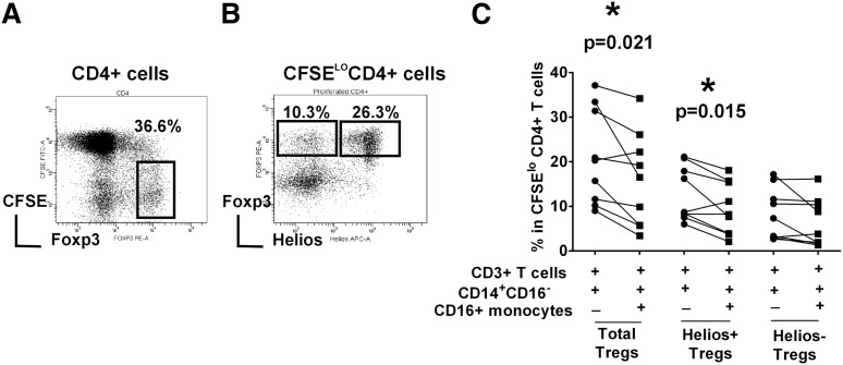
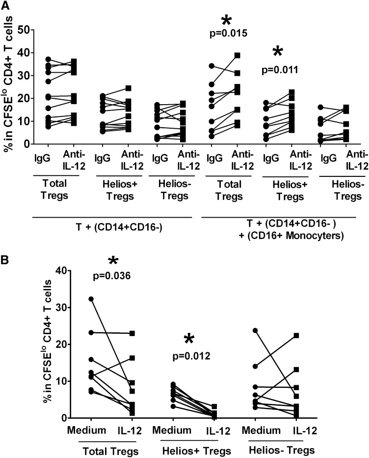
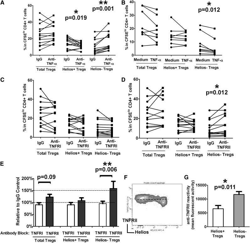
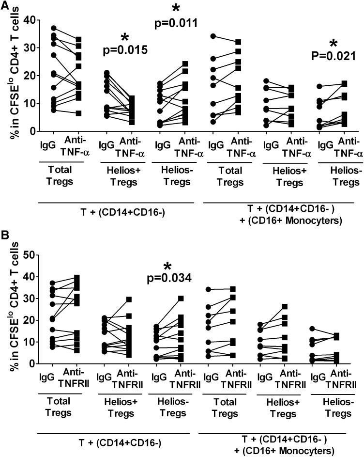
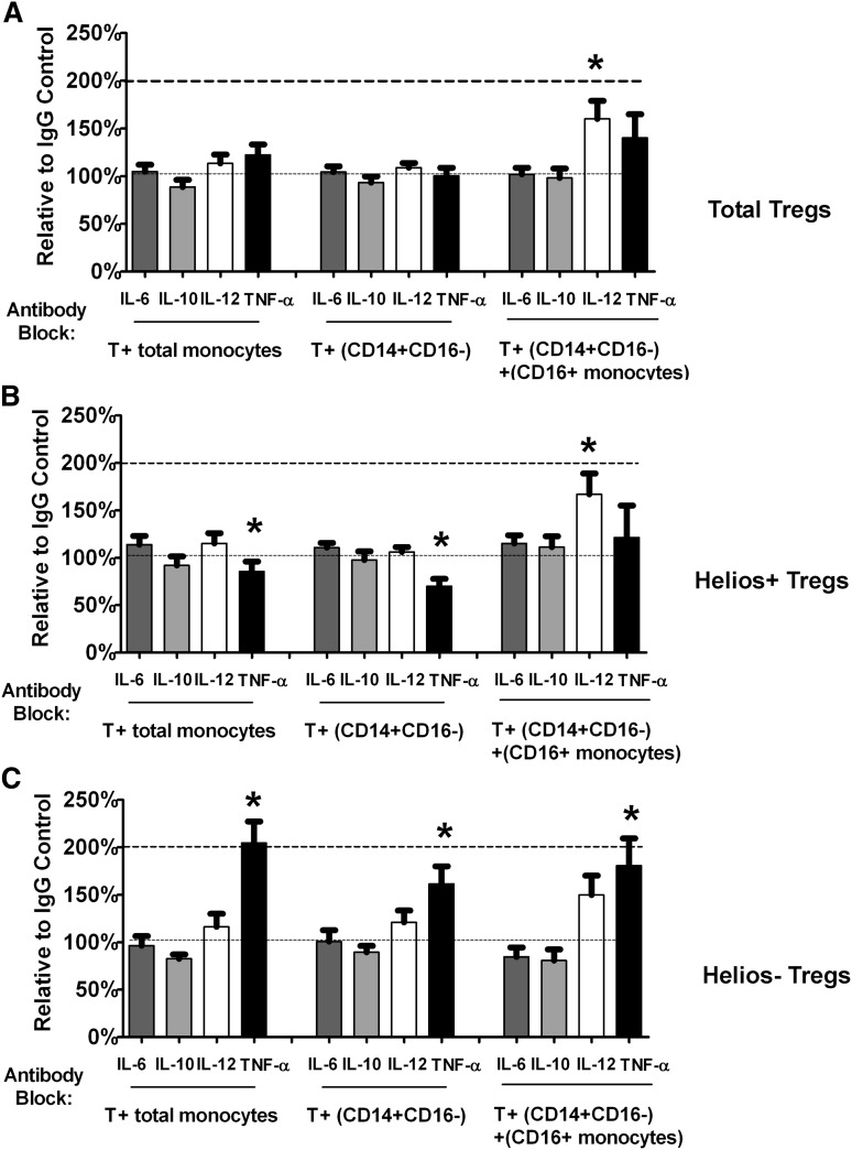
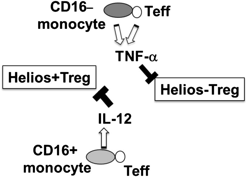
Foxp3(+) regulatory T cells (Tregs) play a pivotal role in control of autoimmunity and pathological immune responses. Helios, the Ikarus family transcription factor, binds to the Foxp3 promoter, stabilizing its expression, and is expressed in 70% of peripheral Tregs of healthy individuals. This frequency is altered during malignancy, infection, and autoimmunity, although the mechanisms that control proliferation and relative numbers of Helios(+/-) Tregs remain largely unknown. Using a T-cell-monocyte in vitro stimulation assay, we now show that proliferation of Helios(+) Tregs is inhibited by CD16(+) monocyte subset. Antibody blocking with anti-interleukin (IL)-12 reversed this inhibition, whereas addition of IL-12 suppressed Helios(+) Treg expansion, indicating that CD16(+) monocyte control of Helios(+) Treg numbers is mediated through IL-12. In contrast, proliferation of Helios(-) Tregs, which express higher levels of tumor necrosis factor receptor II (TNFRII), was suppressed by TNF-α, whereas anti-TNF-α and anti-TNFRII reversed the inhibition. CD16(-) monocyte subset was mainly responsible for TNF-α-mediated control of Helios(-) Treg expansion. Altogether, these data suggest a differential role for monocyte subsets in control of Helios(+/-) Treg development that is mediated by distinct inflammatory cytokines. These data may have important implications for understanding the pathogenesis as well as control of chronic inflammatory and autoimmune diseases.


Figures

Figure 1
Figure 1
Helios+/− Treg proliferation is differentially controlled by monocyte subsets. Autologous CD3+ T cells, CD14+CD16- and CD16+ monocyte subsets were purified from PBMCs of healthy volunteer controls. T cells were CFSE-labeled and co-cultured with CD14+CD16 cells with or without CD16+ monocytes (as indicated by + and −) in the presence of anti-CD3 for 7 days. (A) The gating strategy to analyze the frequency of Foxp3hi in divided (CFSElo) CD4+ subset is shown. (B) Representative dot plot of Helios and Foxp3 expression gated on divided CFSElo CD4+ cells. Gating strategy for cells expressing Foxp3hiHelios+ and Foxp3hiHelios is indicated. (C) The percentage of Foxp3hi in CFSElo CD4+ subset (“Total Tregs”) as well as Foxp3hiHelios+ (“Helios+ Tregs”) and Foxp3hiHelios (“Helios Tregs”) before and after addition of CD16+ monocytes is shown. The P value was calculated by paired t-test and indicates that addition of CD16+ cells decreases Treg development in healthy normal volunteers (P = .021), as per our previously published data, as well as Helios+ Treg development (P = .015), but has a less obvious (not statistically significantly) effect on Helios Treg proliferation.
Figure 2
Figure 2
Helios+ Treg proliferation is inhibited by CD16+ monocytes through IL-12. (A) Purified T cells were co-cultured with purified autologous CD14+CD16 cells without or with CD16+ monocytes in the presence of neutralizing anti–IL-12 or isotype control (“IgG”) antibodies, and after 7 days of stimulation with anti-CD3, the frequencies of Foxp3hi (“Total Tregs”) as well as Foxp3hiHelios+ (“Helios+ Tregs”) and Foxp3hiHelios (“Helios Tregs”) in CFSEloCD4+ subset were analyzed. The P value was calculated by paired t-test comparing the effects of treatment with anti–IL-12 relative to IgG control. In the presence of CD16+ cells, IL-12 neutralization increased total Treg and Helios+ Treg subset proliferation, but not the proliferation of Helios Treg, indicating that CD16+ monocytes inhibit Helios+ Treg development through IL-12. (B) IL-12 was added at the start of the co-cultures of T cells and monocytes, which were stimulated with anti-CD3 for 7 days. Treg (total, Helios+, and Helios) proliferation was then compared in the absence (“Medium”) or presence (“IL-12”) of cytokine by paired t-test. The calculated P values indicate that total Treg and Helios+, but not Helios, Treg proliferation, are inhibited by IL-12.
Figure 3
Figure 3
TNF-α inhibits Helios Treg proliferation through TNFRII. (A) Purified T cells were co-cultured with purified monocytes in the presence of neutralizing anti–TNF-α antibody or an isotype control (“IgG”) antibody, and after 7 days of stimulation with anti-CD3, the frequencies of Foxp3hi (“Total Tregs”) as well as Foxp3hiHelios+ (“Helios+ Tregs”) and Foxp3hiHelios (“Helios Tregs”) in theCFSElo CD4+ subset were analyzed. The P value was calculated by paired t-test comparing the effects of treatment with anti–TNF-α relative to IgG control and indicates that Helios+ Treg proliferation is reduced, whereas Helios Treg proliferation is increased when TNF-α is neutralized through anti–TNF-α. (B) TNF-α was added at the start of the co-cultures of T cells and monocytes stimulated with anti-CD3 for 7 days. Treg (total, Helios+, and Helios) proliferation was then compared in the absence (“Medium”) or presence (“TNF-α”) of cytokine by paired t-test. The calculated P values indicate that only Helios Treg proliferation is targeted by TNF-α, causing a decrease in proliferation of this subset. (C) Co-cultures of T cells and monocytes were treated with anti-TNFRI or (D) anti-TNFRII and stimulated with anti-CD3 for 7 days. Treg (total, Helios+, and Helios) proliferation was then compared with IgG control antibody by paired t-test. The calculated P values indicate that only blocking with anti-TNFRII affects proliferation of Helios Tregs. (E) Percent increase in the numbers of Tregs (total, Helios+, and Helios) in the presence of anti-TNFRI and anti-TNFRII relative to IgG control is shown for the studies presented in (C) and (D) and is consistent with a role for TNFRII, but not TNFRI, in Helios Treg development. (F) Representative dot plot of TNFRII and Helios expression in divided Foxp3hi Tregs, showing surface expression levels of TNFRII on Helios+/− Tregs. (G) Relative mean fluorescence intensity analysis of TNFRII expression on Helios+/− Tregs is shown, indicating that Helios Tregs express higher levels of TNFRII compared with Helios+ Tregs.
Figure 4
Figure 4
CD16 monocytes control TNF-α–mediated Helios Treg proliferation. Purified T cells were co-cultured with purified autologous CD14+CD16 cells without or with CD16+ monocytes in the presence of neutralizing (A) anti–TNF-α or (B) anti-TNFRII, and after 7 days of stimulation with anti-CD3, the frequencies of Foxp3hi (“Total Tregs”) as well as Foxp3hiHelios+ (“Helios+ Tregs”) and Foxp3hiHelios (“Helios Tregs”) in the CFSElo CD4+ subset were analyzed. The P value was calculated by paired t-test comparing the effects of treatment with neutralization antibody relative to IgG control. In co-cultures without CD16+ cells, anti–TNF-α treatment decreased Helios+ proliferation but increased Helios Treg expansion, similar to data obtained when total monocytes were used. In the presence of CD16+ monocytes, Helios Tregs were expanded relative to IgG control. (B) The same assays were performed as in (A) except that for TNF-α blockade, anti-TNFRII was used instead of anti–TNF-α. In co-cultures without CD16+ cells, anti-TNFRII treatment only affected Helios Treg proliferation as indicated by P values, similar to data obtained when total monocytes (Figure 3A) were used.
Figure 5
Figure 5
Helios+/− Treg numbers are not affected by antibody blocking with anti– IL-6 or anti–IL-10 in T cell–monocyte co-cultures. Relative change compared with IgG control in (A) total Treg (“Total Tregs”) as well as (B) Helios+ (“Helios+ Tregs”) and (C) Helios (“Helios Tregs”) Treg numbers in T cell co-cultured with total monocytes or monocyte subsets (CD14+CD16 cells without or with CD16+ monocytes) treated with neutralizing antibodies to IL-6 and IL-10 are shown. For comparison, the relative change in Treg numbers after treatment with anti–IL-12 and anti-TNF-α is also shown.
Figure 6
Figure 6
Hypothetical model of differential regulation of Helios+/− Treg development by monocyte subsets. Interaction of T effector (Teff) with CD16 monocyte subset results in expression of TNF-α, which in turn inhibits Helios Treg development, whereas T cell–CD16+ monocyte interactions trigger IL-12 expression, which inhibits Helios+ Treg proliferative responses.

Similar articles

Cited by

References

    1. Sakaguchi S, Miyara M, Costantino CM, et al. FOXP3+ regulatory T cells in the human immune system. Nat Rev Immunol. 2010;10(7):490–500. - PubMed
    1. Fu W, Ergun A, Lu T, et al. A multiply redundant genetic switch ‘locks in’ the transcriptional signature of regulatory T cells. Nat Immunol. 2012;13(10):972–980. - PMC - PubMed
    1. Getnet D, Grosso JF, Goldberg MV, et al. A role for the transcription factor Helios in human CD4(+)CD25(+) regulatory T cells. Mol Immunol. 2010;47(7-8):1595–1600. - PMC - PubMed
    1. Kim YC, Bhairavabhotla R, Yoon J, et al. Oligodeoxynucleotides stabilize Helios-expressing Foxp3+ human T regulatory cells during in vitro expansion. Blood. 2012;119(12):2810–2818. - PMC - PubMed
    1. Thornton AM, Korty PE, Tran DQ, et al. Expression of Helios, an Ikaros transcription factor family member, differentiates thymic-derived from peripherally induced Foxp3+ T regulatory cells. J Immunol. 2010;184(7):3433–3441. - PMC - PubMed

Publication types

MeSH terms