. 2013:2013:490476.
doi: 10.1155/2013/490476. Epub 2013 Jan 14.

Omega-3 polyunsaturated Fatty acids enhance neuronal differentiation in cultured rat neural stem cells

Affiliations

Omega-3 polyunsaturated Fatty acids enhance neuronal differentiation in cultured rat neural stem cells

Masanori Katakura et al. Stem Cells Int. 2013.

Abstract

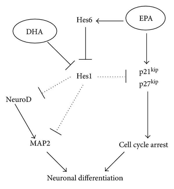
Polyunsaturated fatty acids (PUFAs) can induce neurogenesis and recovery from brain diseases. However, the exact mechanisms of the beneficial effects of PUFAs have not been conclusively described. We recently reported that docosahexaenoic acid (DHA) induced neuronal differentiation by decreasing Hes1 expression and increasing p27(kip1) expression, which causes cell cycle arrest in neural stem cells (NSCs). In the present study, we examined the effect of eicosapentaenoic acid (EPA) and arachidonic acid (AA) on differentiation, expression of basic helix-loop-helix transcription factors (Hes1, Hes6, and NeuroD), and the cell cycle of cultured NSCs. EPA also increased mRNA levels of Hes1, an inhibitor of neuronal differentiation, Hes6, an inhibitor of Hes1, NeuroD, and Map2 mRNA and Tuj-1-positive cells (a neuronal marker), indicating that EPA induced neuronal differentiation. EPA increased the mRNA levels of p21(cip1) and p27(kip1), a cyclin-dependent kinase inhibitor, which indicated that EPA induced cell cycle arrest. Treatment with AA decreased Hes1 mRNA but did not affect NeuroD and Map2 mRNA levels. Furthermore, AA did not affect the number of Tuj-1-positive cells or cell cycle progression. These results indicated that EPA could be involved in neuronal differentiation by mechanisms alternative to those of DHA, whereas AA did not affect neuronal differentiation in NSCs.


Figures

Figure 1
Figure 1
Immunofluorescence images of differentiated neural stem cells (NSCs) stained with Tuj-1 (a neuron marker, (a)–(d)) or GFAP (an astrocyte marker, (e)–(h)) and PI (nuclei) in control (0.01% BSA treated, (a) and (e)), DHA (1 μM, (b) and (f)), EPA (1 μM, (c) and (g)), and AA (1 μM, (d) and (h)) groups on treatment day 4. White bar indicates 10 μm.
Figure 2
Figure 2
Quantification of Tuj-1 (a) or GFAP (b) positive cells in control, DHA, EPA, and AA groups after 2 (white), 4 (gray), and 7 (black) days. Values are presented as means ± SE for five experiments. Statistical analyses were performed by ANOVA followed by Dunnett's test. #,$,∗ P < 0.05 versus control group.
Figure 3
Figure 3
Effects of PUFA on mRNA expression levels of bHLH transcription factors. The values are expressed as the mean ± SE of the fold increase in the ratio of each gene/GAPDH, with the value of the control group (Day 1) taken as 1.0. Statistical analysis was performed by ANOVA followed by Dunnett's test. *P < 0.05 versus control group.
Figure 4
Figure 4
Cell cycle analysis in NSCs after treatment with or without PUFAs. Incorporation of BrdU and total DNA content (7-amino-actinomycin D, 7-AAD) in cells treated with BSA (a), 1 μM DHA (b), EPA (c), or AA (d) were measured by flow cytometry. The cell ycle phase was determined by counting each gate. The percentage of cells distributed in each phase of the cell cycle is shown: G0/G1 phase (e), S phase (f), and G2/M phase (g). Values are means ± SE of three independent experiments. *P < 0.05 versus control group. FITC-BrdU is fluorescein isothiocyanate-conjugated anti BrdU antibody.
Figure 5
Figure 5
Effects of PUFAs on mRNA expression levels of p21cip1 and p27kip1. The values are expressed as the means ± SE of the fold increase in the ratio of each gene versus GAPDH, with the value of the control group (day 1) taken as 1.0. Statistical analysis was performed by ANOVA followed by Dunnett's test. *P < 0.05 versus control group.
Figure 6
Figure 6
Mechanisms of omega-3PUFA-induced neuronal differentiation in NSCs.

References

    1. Perica MM, Delaš I. Essential fatty acids and psychiatric disorders. Nutrition in Clinical Practice. 2011;26(4):409–425. - PubMed
    1. DeCarolis NA, Eisch AJ. Hippocampal neurogenesis as a target for the treatment of mental illness: a critical evaluation. Neuropharmacology. 2010;58(6):884–893. - PMC - PubMed
    1. Van Kampen JM, Eckman CB. Agonist-induced restoration of hippocampal neurogenesis and cognitive improvement in a model of cholinergic denervation. Neuropharmacology. 2010;58(6):921–929. - PMC - PubMed
    1. Thompson A, Boekhoorn K, Van Dam AM, Lucassen PJ. Changes in adult neurogenesis in neurodegenerative diseases: cause or consequence? Genes, Brain and Behavior. 2008;7(1):28–42. - PubMed
    1. Kawakita E, Hashimoto M, Shido O. Docosahexaenoic acid promotes neurogenesis in vitro and in vivo. Neuroscience. 2006;139(3):991–997. - PubMed

LinkOut - more resources