. 2013;8(1):e54631.
doi: 10.1371/journal.pone.0054631. Epub 2013 Jan 24.

Baculovirus superinfection: a probable restriction factor on the surface display of proteins for library screening

Affiliations

Baculovirus superinfection: a probable restriction factor on the surface display of proteins for library screening

Xiaodong Xu et al. PLoS One. 2013.

Abstract

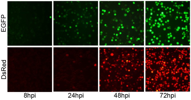
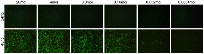
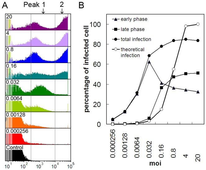
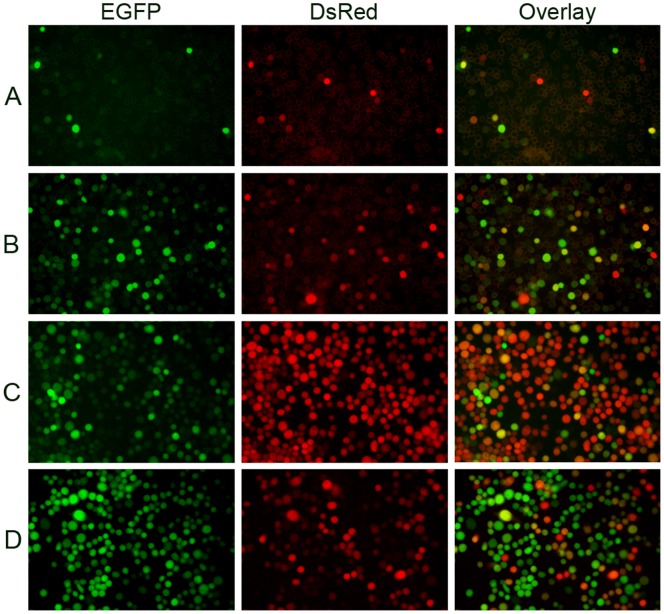
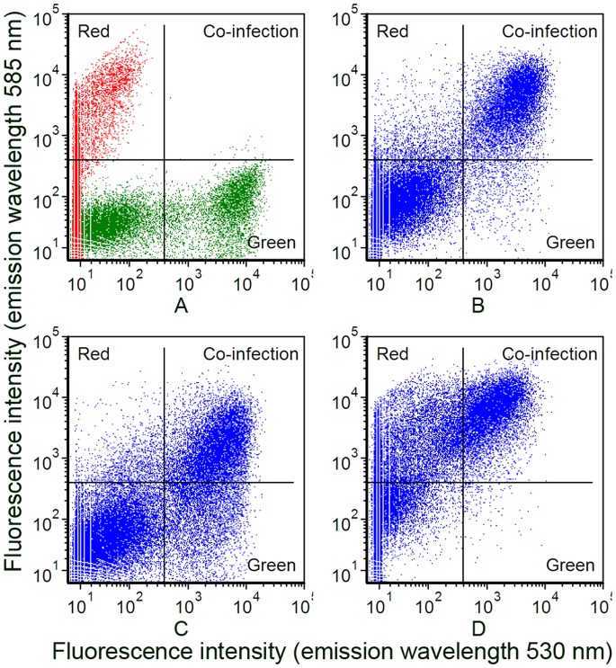
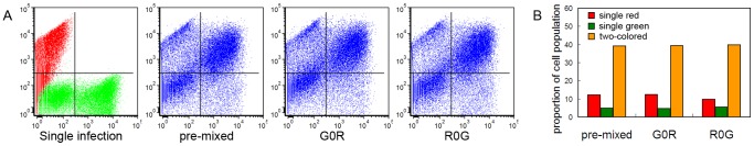
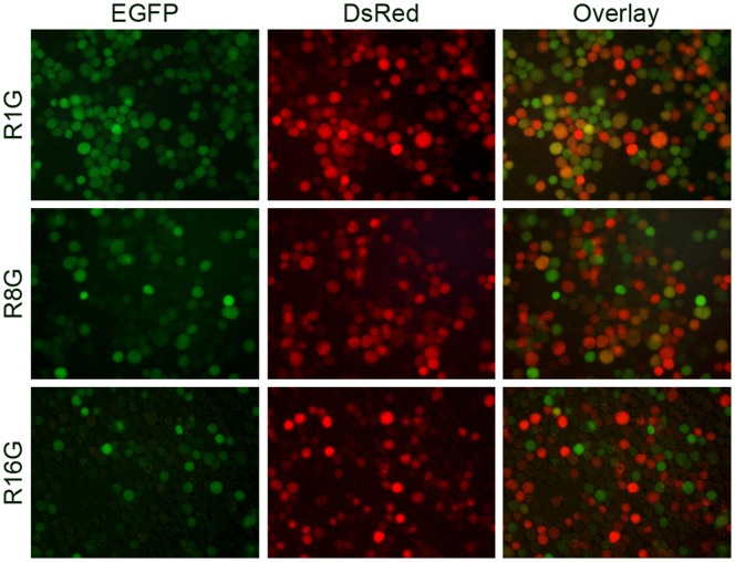
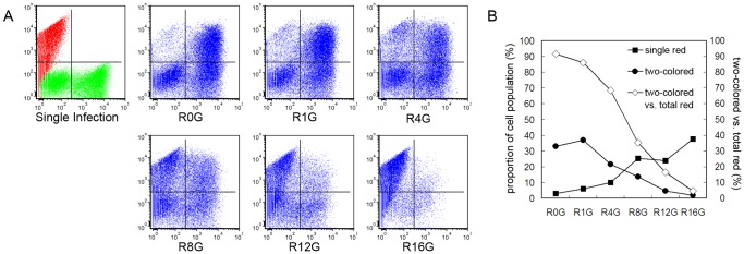
In addition to the expression of recombinant proteins, baculoviruses have been developed as a platform for the display of complex eukaryotic proteins on the surface of virus particles or infected insect cells. Surface display has been used extensively for antigen presentation and targeted gene delivery but is also a candidate for the display of protein libraries for molecular screening. However, although baculovirus gene libraries can be efficiently expressed and displayed on the surface of insect cells, target gene selection is inefficient probably due to super-infection which gives rise to cells expressing more than one protein. In this report baculovirus superinfection of Sf9 cells has been investigated by the use of two recombinant multiple nucleopolyhedrovirus carrying green or red fluorescent proteins under the control of both early and late promoters (vAcBacGFP and vAcBacDsRed). The reporter gene expression was detected 8 hours after the infection of vAcBacGFP and cells in early and late phases of infection could be distinguished by the fluorescence intensity of the expressed protein. Simultaneous infection with vAcBacGFP and vAcBacDsRed viruses each at 0.5 MOI resulted in 80% of infected cells co-expressing the two fluorescent proteins at 48 hours post infection (hpi), and subsequent infection with the two viruses resulted in similar co-infection rate. Most Sf9 cells were re-infectable within the first several hours post infection, but the re-infection rate then decreased to a very low level by 16 hpi. Our data demonstrate that Sf9 cells were easily super-infectable during baculovirus infection, and super-infection could occur simultaneously at the time of the primary infection or subsequently during secondary infection by progeny viruses. The efficiency of super-infection may explain the difficulties of baculovirus display library screening but would benefit the production of complex proteins requiring co-expression of multiple polypeptides.


Conflict of interest statement

Competing Interests: The authors have declared that no competing interests exist.

Figures

Figure 1
Figure 1. Time course for the expression of fluorescent proteins.
Sf9 cells were infected with recombinant AcMNPVs carrying fluorescent marker genes at 2 MOI. Images were taken at 8, 24, 48 and 72 hpi, using fluorescent microscope. EGFP: cells infected with vAcBacGFP; DsRed: cells infected with vAcBacDsRed.
Figure 2
Figure 2. Microscopic analysis of the infection efficiency with vAcBacGFP at different multiplicities of infection.
Sf9 cells were infected with 5 times serially diluted vAcBacGFP viruses, starting from an MOI of 20. The infection was observed and recorded using fluorescent microscope at 24 and 48 hpi.
Figure 3
Figure 3. Flow cytometry analysis of cells infected with vAcBacGFP at different multiplicities of infection at 48 hpi.
Sf9 cells were infected with 5 times serially diluted vAcBacGFP viruses, starting from an MOI of 20. A. Fluorescence histograms. From top to bottom: cells infected at 20, 4, 0.8, 0.16, 0.032, 0.0064, 0.00128, 0.000256 MOI, and uninfected cells as control. B. Comparison of the infection efficiency at different multiplicities of infection. Curves were drawn from the flow cytometry data. Early phase: fraction of cell population in peak 1; late phase: fraction of cell population in peak 2; total infection: fraction of cell population in peak 1 plus peak 2; theoretical infection: theoretical infection level based on the Poisson distribution.
Figure 4
Figure 4. Microscopic analysis of Sf9 cells co-infected with vAcBacGFP and vAcBacDsRed.
Sf9 cells were simultaneously infected with vAcBacGFP and vAcBacDsRed viruses. Protein expression was observed 48 hpi. Many cells were found co-expressing red and green fluorescent proteins even at low multiplicity. A. Cells were infected with 0.05 MOI of each virus. B. Cells were infected with 0.5 MOI of each virus. C. Cells were infected with vAcBacGFP and vAcBacDsRed at a ratio of 1∶4 respectively. D. Cells were infected with vAcBacGFP and vAcBacDsRed at the ratio of 4∶1 respectively.
Figure 5
Figure 5. Detection of cell populations co-expressing EGFP and DsRed by flow cytometry.
A. Individually infected cells. Sf9 cells individually infected with 1 MOI of vAcBacGFP and vAcBacDsRed were mixed at 48 hpi and used as the reference to gate the two-colored and single-colored cell subsets. B. Cells co-infected with vAcBacGFP and vAcBacDsRed at the MOI of 0.5 each. C. Cells co-infected with 0.8 MOI of vAcBacGFP and 0.2 MOI of vAcBacDsRed. D. Cells co-infected with 0.2 MOI of vAcBacGFP and 0.8 MOI of vAcBacDsRed.
Figure 6
Figure 6. Comparison of cell infection using pre-mixed vAcBacGFP and vAcBacDsRed with infections using sequentially added viruses.
A. Flow cytometry data for protein expression at 2 days post infection. Single infection: Sf9 cells were infected individually with vAcBacGFP and vAcBacDsRed then mixed and used as the reference to gate the two-colored and single-colored cell subsets; Pre-mixed: vAcBacGFP and vAcBacDsRed viruses were mixed at a ration of 1∶1, stored at 4°C for 1 day and then used to infect Sf9 cells; G0R: 1 MOI of vAcBacDsRed was applied to Sf9 cell monolayer, mixed well and held for 1 min, before adding 1 MOI of vAcBacGFP; R0G: 1 MOI of vAcBacGFP was applied to Sf9 cell monolayer, mixed well and held for 1 min, before adding 1 MOI of vAcBacDsRed. B. Comparison of the fluorescent cell populations. Bars reflect the flow cytometry data shown in A.
Figure 7
Figure 7. Re-infection of Sf9 cells with homologous baculoviruses.
Sf9 cells were primarily infected with 1 MOI of vAcBacDsRed, and subsequently infected by 1 MOI of vAcBacGFP at an interval of 1 (top panel), 8 (middle panel) or 16 (bottom panel) hours. Protein expression was monitored 48 hpi of vAcBacDsRed by fluorescent microscopy.
Figure 8
Figure 8. Flow cytometry analysis of Sf9 cells re-infected with homologous baculoviruses.
Sf9 cells were primarily infected with 2 MOI of vAcBacDsRed, and subsequently infected by 4 MOI of vAcBacGFP at an interval of 0 (R0G), 1 (R1G), 4 (R4G), 8 (R8G), 12 (R12G) or 16 (R16G) hours. A. Protein expression was analyzed at 48 hpi of infection by vAcBacDsRed. Single infection: Sf9 cells were infected individually with vAcBacGFP and vAcBacDsRed then mixed at 48 hpi and used as the reference to gate the two-colored and single-colored cell subsets. B. Comparison of the fluorescent cell populations at 2 days post the first virus infection. Curves were drawn from the flow cytometry data shown in A. C. Comparison of the fluorescent cell populations at 3 days post the first virus infection.

References

    1. Hitchman RB, Possee RD, King LA (2009) Baculovirus Expression Systems for Recombinant Protein Production in Insect Cells. Recent Patents on Biotechnology 3: 46–54. - PubMed
    1. Makela AR, Oker-Blom C (2008) The Baculovirus Display Technology-An Evolving Instrument for Molecular Screening and Drug Delivery. Combinatorial Chemistry & High Throughput Screening 11: 86–98. - PubMed
    1. James DC, Freedman RB, Hoare M, Ogonah OW, Rooney BC, et al. (1995) N-Glycosylation of recombinant human interferon-gamma produced in different animal expression systems. Biotechnology (NY) 13: 592–596. - PubMed
    1. Hericourt F, Blanc S, Redeker V, Jupin I (2000) Evidence for phosphorylation and ubiquitinylation of the turnip yellow mosaic virus RNA-dependent RNA polymerase domain expressed in a baculovirus-insect cell system. Biochem J 349: 417–425. - PMC - PubMed
    1. Hodder AN, Crewther PE, Matthew ML, Reid GE, Moritz RL, et al. (1996) The disulfide bond structure of Plasmodium apical membrane antigen-1. J Biol Chem 271: 29446–29452. - PubMed

Publication types