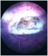
Bladder schwannoma -- a case presentation
- PMID: 23365700
- PMCID: PMC3557122
- DOI: 10.3941/jrcr.v6i12.1232
Bladder schwannoma -- a case presentation
Abstract
Bladder schwannomas are exceedingly rare, benign or malignant, nerve sheath tumors that are most often discovered in patients with a known diagnosis of Neurofibromatosis type 1 (NF1). A few sporadic case reports of bladder schwannoma have been published in urologic, obstetric/gynecologic, and pathologic journals. However, this is the first case report in the radiologic literature where computed tomography imaging and radiology-specific descriptions are discussed. Furthermore, the patient presented in this case is only the fifth published patient without NF1 to be diagnosed with a bladder schwannoma, to the best of our knowledge.
Keywords: Bladder mass; bladder schwannoma; neurofibromatosis; schwannoma.
Figures


References
-
- Wong-You-Cheong JJ, Woodward PJ, Manning MA, Sesterhenn IA. Neoplasms of the urinary bladder: radiologic-pathologic correlation. Radiographics. 2006;26(2):553–80. - PubMed
-
- Murphey MD, Smith WS, Smith SE, Kransdorf MJ, Temple HT. Imaging of musculoskeletal neurogenic tumors: radiologic-pathologic correlation. Radiographics. 1999;19(5):1253–80. - PubMed
-
- Robert PE, Smith JB, Sakr W, Pierce JM. Malignant peripheral nerve sheath tumor (malignant schwannoma) of urinary bladder in von Recklinghausen neurofibromatosis. Urology. 1991;38:473–476. - PubMed
-
- Cummings JM, Wehry MA, Parra RO, Levy BK. Schwannoma of the Urinary Bladder: A Case Report. International Journal of Urology. 1998;38:496–497. - PubMed
-
- Gafson I, Rosenbaum T, Kubba F, Meis JM, Gordon AD. Schwannoma of the bladder: A rare pelvic tumour. Journal of Obstetrics and Gynecology. 2008;28(2):241–254. - PubMed
Publication types
MeSH terms
LinkOut - more resources
Full Text Sources
Medical
Research Materials
Miscellaneous

