Review
. 2012 Dec;6(12):32-42.
doi: 10.3941/jrcr.v6i12.1241. Epub 2012 Dec 1.

Chondrosarcoma in childhood: the radiologic and clinical conundrum

Affiliations
Review

Chondrosarcoma in childhood: the radiologic and clinical conundrum

Susan M Mosier et al. J Radiol Case Rep. 2012 Dec.

Abstract

Less than 10% of chondrosarcomas occur in children. In addition, as little as 0.5% of low-grade chondrosarcomas arise secondarily from benign chondroid lesions. The presence of focal pain is often used to crudely distinguish a chondrosarcoma (which is usually managed with wide surgical excision), from a benign chondroid lesion (which can be followed by clinical exams and imaging surveillance). Given the difficulty of localizing pain in the pediatric population, initial radiology findings and short-interval follow-up, both imaging and clinical, are critical to accurately differentiate a chondrosarcoma from a benign chondroid lesion. To our knowledge, no case in the literature discusses a chondrosarcoma possibly arising secondarily from an enchondroma in a pediatric patient. We present a clinicopathologic and radiology review of conventional chondrosarcomas. We also attempt to further the understanding of how to manage a chondroid lesion in the pediatric patient with only vague or bilateral complaints of pain.

Keywords: bone tumor; chondrosarcoma; pediatric; pediatric chondrosarcoma.


Figures

Figure 1
Figure 1
This is a ten year old male who would be diagnosed 2.75 years later with a low-grade chondrosarcoma. Frog-legged lateral (a) and coned-down frontal (b) radiographs of the left hip demonstrate a faint, round, 8–9 mm lesion (arrowhead) located centrally within the proximal femur. It has a thin sclerotic rim and no evidence of cortical involvement.
Figure 2
Figure 2
This is a 10 year-old male who would be diagnosed 2.75 years later with a low-grade chondrosarcoma. This MR of the left hip took place 3 weeks after the previous radiographs. It demonstrates a round, enhancing, 8–9mm lesion (arrowheads) located centrally in the proximal left femur that is T2WI and STIR hyperintense, and T1WI hypointense. The Philips 1.5 T MR sequences depicted include: Axial STIR (a - top left; supine, TR 3400, TE 81, ST 3), axial T1WI (b - top middle; TR 657, TE 24, ST 3), axial magnified post-contrast T1WI fat-sat (c - top right; 10 cc Magnevist, TR 855, TE 24, ST 3), coronal STIR (d - bottom left; TR 4420, TE 40, ST 2), coronal T1WI (e - bottom middle; TR 604, TE 25, ST 2), and coronal magnified post-contrast T1WI fat-sat (f - bottom right; 10 cc Magnevist, TR 767, TE 25, ST 2)
Figure 3
Figure 3
This is a 10 year-old male who would be diagnosed 2.75 years later with a low-grade chondrosarcoma. Lower extremity, 3 hour delayed PA and AP planar images of a 99Tc-HDP bone scan (a few days after the previous MRI) demonstrate normal musculoskeletal activity without abnormal focal uptake in the proximal left femur (area of concern).
Figure 4
Figure 4
Our patient, now a 12 year-old male, was diagnosed with a low-grade chondrosarcoma a few days after these radiographs. Frontal (a) and frog-legged lateral (b) radiographs of the left hip demonstrate a closed, moderately displaced, pathologic fracture through the [now] lytic lesion (star) in the proximal left femur which has grown to 3.5 cm in maximum transverse dimension. No obvious tumor matrix is visualized.
Figure 5
Figure 5
This is a 12 year-old male who was diagnosed with a low-grade chondrosarcoma a few days after this CT. Oblique sagittal 3D reconstructions (a), coronal (b), and axial (c and d) CT images (1 day after the above radiographs) demonstrate a fracture through the 3.5 cm, hypodense mass centered in the proximal femur. The axial cut superior to the fracture (d) demonstrates endosteal scalloping. The 64 slice CT exam was performed supine with a kVp of 120, mA of 120, ST of 5 for the axial, and ST of 1 for the sagittal and coronal.
Figure 6
Figure 6
This is a 12 year-old male who was diagnosed with a low-grade chondrosarcoma a few days after this MRI. The MR images demonstrate a round, 3.5 cm mass with lobular borders which cause endosteal scalloping greater than 2/3rds the width of the adjacent cortex. It is a STIR hyperintense (bright) mass (star) with extensive adjacent edema (arrowhead). Post-contrast images demonstrate peripheral (c - arrowhead) and central nodular (d - star) enhancement. There is a fracture through the involved proximal femur at the inferior aspect of the mass (d). The Philips 1.5T MR magnet images depicted include: Coronal STIR MRI (a - left; TR 3858, TE 15, ST 5), coronal post-contrast T1WI with fat-suppression (b - middle left; 10 cc Magnevist, TR 715, TE 15, ST 2.91), axial post-contrast T1WI with fat-suppression through the fracture slightly inferior to the mass (c - middle right; 10 cc Magnevist, TR 630, TE 10, ST 3), and axial post-contrast T1WI with fat-suppression (d) through the superior aspect of the mass.
Figure 7
Figure 7
This is a 12 year-old male who was diagnosed with a low-grade chondrosarcoma upon interpretation of this biopsy. Low-powered magnification (20x) of hematoxylin and eosin stains (a) demonstrate normal bone (arrowhead) adjacent to a piece of well-differentiated, neoplastic cartilage (star). 40× magnification (b) demonstrates bi-nucleated chondrocytes consistent with a low-grade chondrosarcoma (arrowhead).
Figure 8
Figure 8
This is a 12 year-old male who was diagnosed with a low-grade chondrosarcoma upon interpretation of this biopsy. Medium-power magnification (a) hematoxylin and eosin stains demonstrate lobulated neoplastic cartilage becoming more cellular toward the periphery of the tumor (star). High-medium-power magnification (b) demonstrates a very myxoid chondrosarcoma with some mild cytologic atypia (specifically binucleated chondrocytes, arrowhead).
Figure 9
Figure 9
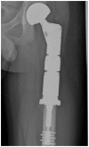
This is a 12 year-old male who underwent resection of a low-grade chondrosarcoma from his proximal left femur with subsequent reconstruction depicted here. An AP radiograph of the proximal left femur demonstrates wide resection of the proximal left femur with interval placement of an intramedullary rod.

References

    1. Murphey MD, et al. From the Archives of AFIP - Imaging of Primary Chondrosarcoma: Radiologic-Pathologic Correlation. RadioGraphics. 2003;23:1245–1278. - PubMed
    1. Hameed M, Dorfman H. Pimary Malignant Bone Tumors - Recent Developments. Semin Diagn Pathol. 2011 Feb;28(1):86–101. - PubMed
    1. Kaste SC, et al. Imaging Pediatric Bone Sarcomas. Radiol Clin North Am. 2011 Jul;49(4):749–65. vi–vii. - PMC - PubMed
    1. Soldatos T, et al. Imaging Features of Chondrosarcoma. J Comput Assist Tomogr. 2011 Jul-Aug;35(4):504–11. - PubMed
    1. Wootton-Gorges SL. MR imaging of primary bone tumors and tumor-like conditions in children. Magn Reson Imaging Clin N Am. 2009 Aug;17(3):469–87. vi. - PubMed

MeSH terms